User talk:Nephron

|
|
Tip: Categorizing images[edit]

Thanks a lot for contributing to the Wikimedia Commons! Here's a tip to make your uploads more useful: Why not add some categories to describe them? This will help more people to find and use them.
Here's how:
1) If you're using the UploadWizard, you can add categories to each file when you describe it. Just click "more options" for the file and add the categories which make sense:

2) You can also pick the file from your list of uploads, edit the file description page, and manually add the category code at the end of the page.
[[Category:Category name]]
For example, if you are uploading a diagram showing the orbits of comets, you add the following code:
[[Category:Astronomical diagrams]][[Category:Comets]]
This will make the diagram show up in the categories "Astronomical diagrams" and "Comets".
When picking categories, try to choose a specific category ("Astronomical diagrams") over a generic one ("Illustrations").
Thanks again for your uploads! More information about categorization can be found in Commons:Categories, and don't hesitate to leave a note on the help desk.BotMultichillT 17:00, 20 September 2009 (UTC)
- Image:Liver reticulin.jpg was uncategorized on 19 September 2009.
- Image:Parathyroid adenoma low mag.jpg was uncategorized on 20 September 2009.
- Image:Parathyroid adenoma intermed mag.jpg was uncategorized on 20 September 2009.
- Image:Parathyroid adenoma high mag.jpg was uncategorized on 20 September 2009.
- Image:Small intestine neuroendocrine tumour high mag cropped.jpg was uncategorized on 23 September 2009.
- Image:Cholestasis high mag.jpg was uncategorized on 26 September 2009.
- Image:Rete testis with seminoma.jpg was uncategorized on 10 June 2010 CategorizationBot (talk) 10:58, 11 June 2010 (UTC)
- Image:Nipple adenoma - low mag.jpg was uncategorized on 6 August 2011 CategorizationBot (talk) 12:59, 7 August 2011 (UTC)
- Image:Nipple adenoma - intermed mag.jpg was uncategorized on 6 August 2011 CategorizationBot (talk) 12:59, 7 August 2011 (UTC)
- Image:Nipple adenoma - very high mag.jpg was uncategorized on 6 August 2011 CategorizationBot (talk) 12:59, 7 August 2011 (UTC)
I was considering using your image of Pheochromocytoma in a presentation, who do I attribute it to?
Hi Nephron,
I would like to use your picture of the Masson's trichrome stained artery cross-section for a talk on the transcriptomic basis of atherosclerosis. Do I have your permission? If so, who do I attribute it to?
Thanks,
Shurjo
Dear Nephron,
My name is James. I am assisting in publishing an ENT review book. We are looking for permission to reprint your image of paraganglioma. Do you have a copy that's at least 300 dpi?
If this is possible, we will include the following credit line and copyright acknowledgement with your reproduction:
Image reprinted with permission from Nephron.
I have attached the image we would like to request permission to use in our book. Please feel free to contact me via email at studiojw@gmail.com
Thank you in advance for all your time and help.
Sincerely,
James
- Hi James:
- If you are referring to the carotid body tumour, I do have the images in higher resolution, but I am not inclined to upload them because they aren't quite in focus. I will re-take the images at some time in the future. What is the time frame of the book? Nephron T|C 00:16, 15 July 2010 (UTC)
- I re-photographed the carotid body tumour (paraganglioma). The images are here: File:Carotid_body_tumour_2_low_mag.jpg, File:Carotid_body_tumour_2_intermed_mag.jpg and File:Carotid_body_tumour_2_high_mag.jpg. Whether they are 300 DPI, depends on how large you make 'em on the page. Nephron T|C 18:24, 17 July 2010 (UTC)
Hi Nephron- My name is Caroline and I work at an ad agency. My coworker especially liked one of your images and we were wondering how to get permission to possibly use any of your images?
- Hi Caroline,
- You don't need my explicit permission -- but you do have to abide by the licensing (GFDL or CC), which is notated in detail for each image (e.g. licensing info an image I created). I briefly described this on my user page. I would be happy to hear where you're going to make use of the image(s) and which image(s)-- though this isn't required by the licensing. Nephron T|C 18:00, 17 July 2010 (UTC)
Hi Nephron, I am an italian high school student and my dream is to be an anatomical pathologist, I want to tell you that I liked each one of your images and I want to thank you because with your images I can "feed" my passion and my interest. --GBMorgagni (talk) 19:04, 20 July 2010 (UTC)
Dear Nephron, this is an advanced-feedback: for a publication (by Ernst Schering Foundation Berlin, Germany) comming out march, 24 2011, I like to use your picture "MI_with_contraction_bands". The book is about "fingerprints..." and completes the same named exhibition the Foundation’s project space, where the performative installations and live laboratories by US-American biomedia artist Paul Vanouse is shown now. Curated by Jens Hauser, the exhibition presents the world premiere of the "Suspect Inversion Center," (SIC) an operational laboratory in which Vanouse creates identical “genetic fingerprints” of criminals and celebrities from his own DNA... Please look here for more information: http://www.scheringstiftung.de/en/project-space/news/2528-paul-vanouse-fingerprints-.html. Thank you for the possibility to use your pictures for free. Kind regards Linda. (For an answer: linda . stanke @ google mail . com)
- Thanks for the quick note! I'd go to the show... but, unfortunately, I'm not currently in Berlin. It would be nice if Vanouse made his this work available on the internet... so I could see it and the derivative of my work. Nephron T|C 05:45, 8 April 2011 (UTC)
use of a photo in a commercial brochure[edit]
Hi,
I work at a cell culture firm and we woudl like to use your photo of Thumus tissue on the cover of our brochure. We are happy to give you the credit in the document. I assume that the contents of the brochure, which will not refer to the photo is aggregated materials and there would not be an unlimited use of our content. Are you OK with that? Terry.taciuk@stemcell.com
- You have to follow a license associated with the image. AFAIK, the GDFL does not prohibit commercial use. Nephron T|C 05:45, 8 April 2011 (UTC)
Do you have similar photos you would be willing to liscence for our commercial use?
Hi,
Are you sure this image is of a normal kidney? To me it looks like there's pretty massive inflammatory infiltrate and quite a bit of cellular debris in the tubules, which would be more suggestive of a transplant rejection. Blahdenoma (talk) 03:31, 20 April 2011 (UTC)
- Describe what it is about in a short sentence. (What does the image show?)
- State the author and the date of creation. If you made it yourself, say so explicitly. If it is from another Wikimedia user, link to the person's local user page. Best to use CommonsHelper.
- If you did not create the file yourself, state the source you got it from.
- Add a copyright tag - images without an appropriate license tag will be deleted.
- Add the image to one or more gallery pages and/or appropriate categories, so it can be found by others. To find out where an image belongs, you can use CommonsSense.
If you copied the file from another wiki, please copy all information given there and say who uploaded it to that wiki. Use CommonsHelper.
It is recommended to use Template:Information to put that information on the description page. Have a look at Template talk:Information for details of the use of this template.
You can edit the description page and change the text. Uploading a new version of the file does not change the description of the file.
Please add as much information as possible. If there is not sufficient information, the file may have to be deleted. For more information, follow the Commons:First steps guide. If you need help or have questions, please ask at the Help desk.
Thank you.This message was added automatically by Nikbot, if you need some help about it, please read the text above again and follow the links in it,if you still need help ask at the ![]() ->Commons:Help desk in any language you like to use. --Nikbot 03:54, 9 November 2011 (UTC)
->Commons:Help desk in any language you like to use. --Nikbot 03:54, 9 November 2011 (UTC)
Hello,
the casts in the picture seem to me PAS positive, so this isn't a myeloma cast nephropathy if you write "Hyaline casts are PAS positive. Myelomatous casts are PAS negative".? --Patho (talk) 09:52, 3 December 2011 (UTC)
This message was added automatically by Nikbot, if you need some help about it please read the text above again and follow the links in it, if you still need help ask at the ![]() → Commons:Help desk in any language you like to use. --Nikbot 05:19, 21 December 2011 (UTC)
→ Commons:Help desk in any language you like to use. --Nikbot 05:19, 21 December 2011 (UTC)
Melanosis coli[edit]
Dear Nephron,
My name is Michael and I would like to use your histological slide on melanosis coli for a chapter in a book I am contributing to about Gastroenterology MCQs. I am looking for permission to reprint your image of histological melanosis coli. Do you have a copy that's at least 300 dpi? I will certainly insert the credit line and copyright acknowledgement. I would be grateful if you could contact me on: michaelfung@gmx.de Kind regards, Michael
- Hi Michael,
- I moved your text to the bottom -- as it is customary to add to the bottom of the page. You are welcome to use any of my images as is -- provided you follow one of the (copyleft) licenses (CC Share-alike or GDFL). Whether a given image is 300 dpi, depends on how big you make the image on the page, and its resolution. Generally, I don't have the images in higher resolution that on the WC. Nephron T|C 04:19, 10 March 2012 (UTC)
Location[edit]
Dear Nephron,
can you give information about the location (organ/tissue) in your fotos? So we/I can better use them in organ specific contexts.
e.g.: File:Aspergillus_-_intermed_mag.jpg
Many thanks for sharing these great fotographs!
--Patho (talk) 10:50, 13 May 2012 (UTC)
Permission for File:RCA atherosclerosis.jpg[edit]
Hi, I wish to use your photo of File:RCA atherosclerosis.jpg in my science book. Please kindly email me at jouehuey.yeoh@pearson.com for any permission clearance I need to know. Thank you.
Joue Huey
- Hi Joue Huey:
- I don't generally do email -- unless it is via the Wikicommons, i.e. you are another Wikicommons user and send me a private message. I don't think the image is one of my better ones, as it has a bad asymmetric vignetting effect. I am planning to redo that image at some point in the future, if I find the time and come across a good case. Above said, you are welcome to use it as long as you follow one of the licences (CC or GFDL). If you are going to use it, I'd appreciated it if you can post a link here to the book/information about the book when it is done. Nephron T|C 17:03, 18 May 2012 (UTC)
Hi Nephron Thank you, I will follow the licenses, I suggest to credit you as User:Nephron @ Wikimedia Commons, CC by-sa. Currently I also wish to use another photo of your File:Small intestine low mag.jpg. I will credit as well. Thank you for sharing all these wonderful photos.
Joue Huey
- I put some more comments on your talk page. Nephron T|C 13:53, 30 May 2012 (UTC)
- Thanks a lot Nephron for the image and also advising me on the usage.
- User talk:Jouehuey 60.54.219.19 01:49, 1 June 2012 (UTC)
Published Reference for Teratoma Image[edit]
Hello Nephron,
I wish to use your teratoma image (http://upload.wikimedia.org/wikipedia/commons/1/11/Teratoma_2_low_mag.jpg) for a presentation of mine, but cannot use the wikipedia citation. Is this image form a published scientific article, or do you have another citation which I could use as a reference?
Thanks! Arthur
Hello Nephron,
for me the epithelium looks unkeratinized, because I see no superificial eosinophilic tinge, no absent nuclei and no keratin lamellae, so why do you think is this not an (unkeratinized) dentigerous cyst? --Patho (talk) 12:12, 27 May 2012 (UTC)
- Describe what it is about in a short sentence. (What does the image show?)
- State the author and the date of creation. If you made it yourself, say so explicitly. If it is from another Wikimedia user, link to the person's local user page. Best to use CommonsHelper.
- If you did not create the file yourself, state the source you got it from.
- Add a copyright tag - images without an appropriate license tag will be deleted.
- Add the image to one or more gallery pages and/or appropriate categories, so it can be found by others. To find out where an image belongs, you can use CommonsSense.
If you copied the file from another wiki, please copy all information given there and say who uploaded it to that wiki. Use CommonsHelper.
It is recommended to use Template:Information to put that information on the description page. Have a look at Template talk:Information for details of the use of this template.
You can edit the description page and change the text. Uploading a new version of the file does not change the description of the file.
Please add as much information as possible. If there is not sufficient information, the file may have to be deleted. For more information, follow the Commons:First steps guide. If you need help or have questions, please ask at the Help desk.
Thank you.This message was added automatically by Nikbot, if you need some help about it, please read the text above again and follow the links in it,if you still need help ask at the ![]() ->Commons:Help desk in any language you like to use. --Nikbot 10:19, 31 May 2012 (UTC)
->Commons:Help desk in any language you like to use. --Nikbot 10:19, 31 May 2012 (UTC)
Permission to use Images of Glycogen Storage Disorder[edit]
Dear Nephron,
I request your kind permission to use images of Glycogen Storage Disorder in my book of General Pathology which will be published shortly. The images will be referenced as per your instructions. I am unable to understand the legal terms used in the webpage. Can you kindly help me. I would appreciate if you can kindly contact me at my e-mail id: drsudhashivaraj@yahoo.com
Thank you Warm Regards Dr. Sudha
- Hello Dr. Sudha:
- Nice to hear you find my images good enough for your book. Details about re-use of images on the Commons are here. Generally, if it is a book, you should (1) give the image credit where the image appears, e.g. Nephron (c) 2007, CC-BY-SA 3.0, and (2) print the full licence (CC-BY-SA or GFDL) in an appendix, such that your readers know they can reuse and distribute the images under the conditions specified in the license. Nephron T|C 19:55, 22 September 2012 (UTC)
Dear Nephron, Thank you. I went through your entire gallery, your images are not only good enough they are very beautiful, i loved them. You have done a wonderful job. Being in a non-academic setup I was having difficulty in finding photographs of rare disorders and tumors. Im glad I found your images. Can I please use a few more of your pictures for the chapter on Cancer Biology. They will all be referenced as per your instructions which I will be sending to my publisher. I will let you know once my book is [published]. Thanks again. Yours Sincerely Sudha
- You are welcome to use any of the images (provided you follow one of the licences). I would be happy to hear from you again when the book is published. You should review the images carefully. I've done my best to take pictures that are representative of what the image is labeled -- but it is possible I didn't get the most representative field. Nephron T|C 13:17, 23 September 2012 (UTC)
Thanks from a student[edit]
Nephron-
Thanks much for your prolific pathology posting. I've been using many of your images in crafting my Pathology reviews for local circulation between med students here at our school. Having your Creative Commons library at my disposal is exceptional as a resource to illustrate the key features of a variety of conditions. Keep up the good work! You're getting many students through to levels of understanding that would be otherwise fraught with a cacaphony of distracting or irrelevant information. Thank-you for keeping our education vividly illustrated.
--StrangeAesculapian
- It is nice to hear that they are appreciated. Nephron T|C 23:20, 14 October 2012 (UTC)
Nephron - Thank you, thank you and thank you. Your top class images in Pathology (I capitalize it because I respect it) have really made Step 1 study a lot easier, esp. with all the eponymous jargon involved. Just like your pictures, things are so much more clearer now. Thank you again.
Ali
TY for upload. Are you able to describe the cellular changes demonstrated? I feel it would be good to include these in the caption or the etiology section. Thanks in advance. 212.183.140.32 12:31, 15 October 2012 (UTC)
- someone from WPMED has supplied this info now. TY again for upload. 212.183.128.144 21:05, 27 October 2012 (UTC)
use of image on a book cover[edit]
Hi Nephron,
may we have your permission to use the photo (File:Small cell lung cancer - cytology.jpg) on the cover of our book? If we have your permission, we will give you credit for the photo. Please feel free to email me at fting.lee@gmail.com
Many thanks in advance, Fengting
- Nice to hear you find the image good enough for your book. Details about re-use of images on the Commons are here. Usually, one puts the image credit where the image appears, e.g. Nephron (c) 2010, CC-BY-SA 3.0, but I think it is customary to put it one the page after the inside cover -- for the cover image. Beyond that, you need to print the full licence (CC-BY-SA or GFDL) in an appendix of the book, such that your readers know they can reuse and distribute the image under the conditions specified in the license. If you can place a link to a picture of the cover... I'd be curious to see it-- though that isn't necessary to use the image. Nephron T|C 04:01, 20 November 2012 (UTC)
Hi Nephron,
thanks for your reply. Can I confirm that you hold the copyright for the image and that we may use it in our cover? Of course, if we do use it, we will cite the source accordingly. I have a draft cover out already but I will require a high-res image file if I'm going to use it for the actual cover. Do you have a high res file for the image? I'll send you the cover via email so that you can have a look at it. My address is fting.lee@gmail.com, please do drop me an email.
Microphotographs showing effects of Radiation[edit]
Dear Nephron, Hi, Im Dr. Sudha and I had written to you few months back requesting permission to use your images in my book of general pathology. The book is progressing well and will be published in 2013. I badly need microphotographs showing the effects of radiation. If you have any can you please give them to me ? I will be extremely grateful to you for the same and they will be licensed according to your instructions. Thank You Warm Regards Sudha
- I don't have a good example of radiation effect -- at least not yet. Nephron T|C 03:40, 4 February 2013 (UTC)
Picture of the Year voting round 1 open[edit]
Dear Wikimedians,
Wikimedia Commons is happy to announce that the 2012 Picture of the Year competition is now open. We're interested in your opinion as to which images qualify to be the Picture of the Year for 2012. Voting is open to established Wikimedia users who meet the following criteria:
- Users must have an account, at any Wikimedia project, which was registered before Tue, 01 Jan 2013 00:00:00 +0000 [UTC].
- This user account must have more than 75 edits on any single Wikimedia project before Tue, 01 Jan 2013 00:00:00 +0000 [UTC]. Please check your account eligibility at the POTY 2012 Contest Eligibility tool.
- Users must vote with an account meeting the above requirements either on Commons or another SUL-related Wikimedia project (for other Wikimedia projects, the account must be attached to the user's Commons account through SUL).
Hundreds of images that have been rated Featured Pictures by the international Wikimedia Commons community in the past year are all entered in this competition. From professional animal and plant shots to breathtaking panoramas and skylines, restorations of historically relevant images, images portraying the world's best architecture, maps, emblems, diagrams created with the most modern technology, and impressive human portraits, Commons features pictures of all flavors.
For your convenience, we have sorted the images into topic categories. Two rounds of voting will be held: In the first round, you can vote for as many images as you like. The first round category winners and the top ten overall will then make it to the final. In the final round, when a limited number of images are left, you must decide on the one image that you want to become the Picture of the Year.
To see the candidate images just go to the POTY 2012 page on Wikimedia Commons
Wikimedia Commons celebrates our featured images of 2012 with this contest. Your votes decide the Picture of the Year, so remember to vote in the first round by January 30, 2013.
Thanks,
the Wikimedia Commons Picture of the Year committee
Delivered by Orbot1 (talk) at 11:12, 19 January 2013 (UTC) - you are receiving this message because you voted last year
Case report of ASPS[edit]
Dear Neprhon,
We would like to use your image of Alveolar Soft Part Sarcoma in a poster presentation that will be given locally as well as nationally. In addition, we would like to ask permissions to use it in a case report manuscript. Please advise us on what the best was is to give copyright/permissions on our poster and manuscript. Thanks. Feel free to email me at studiojw@gmail.com.
Best,
jw
- Hi JW - You are welcome to use my images how you see fit. How to re-use my images is spelled-out on my user page.
- In the context of a case report -- if you're not using the pictures for comparison purposes, it would be more appropriate to use images from the case. I would suggest you contact the pathologist that made the diagnosis. They can probably supply you images and also help you understand the elements of the pathology report that should be in the case report, e.g. confirmatory molecular testing for the characteristic chromosomal translocation, findings from electron microscopy (if done). Nephron T|C 04:48, 24 February 2013 (UTC)
Thanks![edit]
Hi Nephron - I just wanted to thank you for putting your images up, they've really helped with my boards studying!
- I am happy to hear they are being put to use. Good luck on the boards - if you haven't written 'em yet. Nephron T|C 00:09, 13 May 2013 (UTC)
World Cancer Report 2014[edit]
Dear Nephron,
The International Agency for Research on Cancer is currently working on the World Cancer Report 2014. In that context, we would like to use your "High magnification micrograph of ground glass hepatocytes, as seen in a chronic hepatitis B infection with a high viral load".
I noted that your material is licensed under the Attribution-Share Alike 3.0 Unported licence, but would however like to give you an opportunity to tell us if you feel strongly against having your material reproduced in our publication. Additionally, I wanted to check whether the use of your material by the Agency enters your requirements i.e. "images for non-free use". The IARC is part of the World Health Organization (WHO), which is a specialized agency of the United Nations, and operates on a non-profit basis.
Should you agree to grant us permission, you would be credited as follows: "Copyright © 2009 Nephron, Permission is granted to copy, distribute and/or modify this image under the terms of the Attribution-Share Alike 3.0 Unported licence (https://creativecommons.org/licenses/by-sa/3.0/deed.en)."
Looking forward to your reply,
Best regards,
S. Quennehen Editorial clerk - Communications Group International Agency for Research on Cancer
- Hello S.
- I am not sure which image you are referring to. I would suggest this image of GGHs. In any case, you are welcome to use any of my images. If you're going to use them on a web site, I would ask you to link the image on the Wikimedia Commons and my user page. For example:
- "Copyright © 2009 Nephron, Permission is granted to copy, distribute and/or modify this image under the terms of the Attribution-Share Alike 3.0 Unported licence (https://creativecommons.org/licenses/by-sa/3.0/deed.en)."
- Nephron T|C 22:59, 25 June 2013 (UTC)
Thank you for your quick reply. This is indeed the image I was referring to. Since our book will come out in print, I hope it is still acceptable to use the credit line you provided. Your requirements are duly noted, should our publication come out as an e-pub. Thank you for your help, Best, Solene Q - IARC
Ragged Red Fibers[edit]
Hi, I'd like to use your photo of Ragged Red Fibers in a Genetics text book. Please email me to discuss permission and a repro fee. many thanks. jerry(at)pictureresearching.com
- Explicit permission isn't required for usage. Above said, you need to following the licensing terms -- though they aren't very onerous.
- If you want to send money somewhere, I suggest a donation to the Wikipedia Foundation in my pseudonym (Nephron). Nephron T|C 22:29, 10 July 2013 (UTC)
Using your images in my book[edit]
Hi Nephron, I will be really thankful to you if you let me use 2 of your images in a book of Pathology I am about to publish within a few days. I want to attribute you in my book under each image I will use. Please mail me your real name, which will be published in my book. Thank you. Please reply soon.
Thanking you, Prithwiraj Maiti. R.G.Kar Medical College, India. Email Id: prithwiraj2009@yahoo.in
- Hello,
- See Use of the images I have uploaded on my user page for attribution. All the best with your book. Nephron T|C 13:47, 1 September 2013 (UTC)
Copyright status: File:Positive margin with cautery artefact - adenocarcinoma - high mag.jpg[edit]
| This media may be deleted. |
Thanks for uploading File:Positive margin with cautery artefact - adenocarcinoma - high mag.jpg. I notice that the file page either doesn't contain enough information about the license or it contains contradictory information about the license, so the copyright status is unclear.
If you created this file yourself, then you must provide a valid copyright tag. For example, you can tag it with {{self|GFDL|cc-by-sa-all}} to release it under the multi-license GFDL plus Creative Commons Attribution-ShareAlike All-version license or you can tag it with {{PD-self}} to release it into the public domain. (See Commons:Copyright tags for the full list of license tags that you can use.) If you did not create the file yourself or if it is a derivative of another work that is possibly subject to copyright protection, then you must specify where you found it (e.g. usually a link to the web page where you got it), you must provide proof that it has a license that is acceptable for Commons (e.g. usually a link to the terms of use for content from that page), and you must add an appropriate license tag. If you did not create the file yourself and the specific source and license information is not available on the web, you must obtain permission through the VRT system and follow the procedure described there. Note that any unsourced or improperly licensed files will be deleted one week after they have been marked as lacking proper information, as described in criteria for deletion. If you have uploaded other files, please confirm that you have provided the proper information for those files, too. If you have any questions about licenses please ask at Commons:Village pump/Copyright or see our help pages. Thank you. |
JuTa 23:45, 28 September 2013 (UTC)
Picture of the Year 2013 R1 Announcement[edit]
Hi Nephron,
my name is Mike. I'm currently trying to get the LFB-H&E stain to work but failed so far. Do you have a protocol you can provide me with please email me mikeschmidt8@hotmail.com
thanks best regards mike
question[edit]
Hi Nephron,
my name is Mike. I'm currently trying to get the LFB-H&E stain to work but failed so far. Do you have a protocol you can provide me with please email me mikeschmidt8@hotmail.com
thanks best regards mike
Picture of the Year 2013 R2 Announcement[edit]
Round 2 of Picture of the Year 2013 is open![edit]
- ⧼Wikibase-terms/Nephron⧽: Deutsch, Ελληνικά, English, français, magyar, italiano, македонски, 日本語, русский, svenska

Dear Wikimedians,
Wikimedia Commons is happy to announce that the second round of the 2013 Picture of the Year competition is now open. This year will be the eighth edition of the annual Wikimedia Commons photo competition, which recognizes exceptional contributions by users on Wikimedia Commons. Wikimedia users are invited to vote for their favorite images featured on Commons during the last year (2013) to produce a single Picture of the Year.
Hundreds of images that have been rated Featured Pictures by the international Wikimedia Commons community in the past year were entered in this competition. These images include professional animal and plant shots, breathtaking panoramas and skylines, restorations of historical images, photographs portraying the world's best architecture, impressive human portraits, and so much more.
There are two total rounds of voting. In the first round, you voted for as many images as you liked. The top 30 overall and the most popular image in each category have continued to the final. In the final round, you may vote for just one image to become the Picture of the Year.
Round 2 will end on 7 March 2014. https://commons.wikimedia.org/wiki/Special:MyLanguage/Commons:Picture_of_the_Year/2013/Introduction/en Click here to learn more and vote »]
Thanks,
the Wikimedia Commons Picture of the Year committee
You are receiving this message because you voted in the 2013 Picture of the Year contest.
This Picture of the Year vote notification was delivered by MediaWiki message delivery (talk) 19:21, 22 February 2014 (UTC)
Picture of the Year 2013 Results Announcement[edit]
Picture of the Year 2013 Results[edit]
- In other languages: Deutsch, español, français, 日本語, Nederlands, русский, svenska, Türkçe, українська

Dear Nephron,
The 2013 Picture of the Year competition has ended and we are pleased to announce the results: We shattered participation records this year — more people voted in Picture of the Year 2013 than ever before. In both rounds, 4070 different people voted for their favorite images. Additionally, there were more image candidates (featured pictures) in the contest than ever before (962 images total).
- In the first round, 2852 people voted for all 962 files
- In the second round, 2919 people voted for the 50 finalists (the top 30 overall and top 2 in each category)
We congratulate the winners of the contest and thank them for creating these beautiful images and sharing them as freely licensed content:
- 157 people voted for the winner, an image of a lightbulb with the tungsten filament smoking and burning.
- In second place, 155 people voted for an image of "Sviati Hory" (Holy Mountains) National Park in Donetsk Oblast, Ukraine.
- In third place, 131 people voted for an image of a swallow flying and drinking.
Click here to view the top images »
We also sincerely thank to all 4070 voters for participating and we hope you will return for next year's contest in early 2015. We invite you to continue to participate in the Commons community by sharing your work.
Thanks,
the Picture of the Year committee
You are receiving this message because you voted in the 2013 Picture of the Year contest.
Delivered by MediaWiki message delivery (talk) 22:59, 26 March 2014 (UTC)
Permission to print and attribution[edit]
"Como ves" (www.comoves.unam.mx) is a science magazine for high school students and young adults published and subsidized by the National Autonomous University of Mexico. We are currently preparing an article on papanicolau tests and would like to use some of your images to illustrate it. I would like to know if this is possible and if you want to use your user name or any other. Looking forward to hearing from you Yours faithfully
Isabelle Marmasse Assistant Editor imarmase@universum.unam.mx
- Please see Use of the images I have uploaded on my user page. Nephron T|C 00:50, 4 April 2014 (UTC)
Ok, but I believe it might look a little weird. Can I send you a PDF of the article so you can check it?
imarmase@universum.unam.mx
Whipple Disease[edit]
Hey -- I'm looking at your lovely images of Whipple's disease histology, but I'm having a lot of trouble figuring out what I'm looking at -- are all the clear/cloudy cells under the epithelium the foamy macrophages? Are they considered PAS positive, or would those just be the occasional very-red ones? Would you consider making a labeled version of the image for the entry? (I'd whip one up myself, but I'm just a med student, and have very little confidence in my accuracy. :D) Eccentricity (talk) 01:19, 10 April 2014 (UTC)
- I overlooked your post; so, I am replying to it now.
- I only did pictures of a H&E stained slide; I did not have the other stains available to me. PAS is a special stain. "PAS-positive" means the something lights up with it, e.g. some micro-organisms.
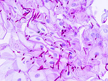
PAS stain showing candida. - Here is a picture showing Whipple's disease with a PAS stain: [2].
_PAS_stain.jpg)
- Tropheryma whipplei (the bug that causes Whipple's disease) is a rod-shaped organisms -- it cannot be seen on the images I took. The organisms are usually in the macrophages -- which fill the lamina propria. Lamina propria macrophages are very abnormal. The histologic differential diagnosis is mycobacterium avium complex (MAC); this is sorted out with stains (acid fast stain to look for MAC, PAS to look for T. whipplei). I hope that helps.
- Nephron T|C 23:54, 19 April 2014 (UTC)
- Oh my! I'm sorry; I just ignored the caption entirely, mistook the strikingly eosinophilic epithelial cells as "red," and assumed I was looking at a PAS slide. Then when I couldn't see any red macrophages, the confusion started. :D The Flickr image clears things up nicely for me.
- I appreciate the patient response, and do hope you keep on raising Wikipedia's game with these beautiful shots!
- --Eccentricity (talk) 01:43, 25 April 2014 (UTC)
Question about "File:Periportal hepatosteatosis"[edit]
Dear Nephron, My name is I, a japanese hepatologist. Thank you for providing beautiful pathologic images to Wikimedia Commons. I'm interested in your file(Periportal Hepatosteatosis) very much. Would you teach me about the differential diagnosis of periportal hepatosteatosis? I think the periportal steatosis is rare phenomenon in NAFLD and mainly present in children. In your differential diagnosis, (e.g. hepatitis C type 3, starvation, total parenteral nutrition) I think it may due to depletion of the VLDL secretion from zone 1. Please tell me the reference paper or the textbook about differential diagnosis (C-TAPES).
Sincerely, I
Hello I: I found the list on http://www.pathconsultddx.com in 2009. Unfortunately, that is no longer available. I did find something about periportal steatosis in children.[3] The classic teaching is that NAFLD is centrilobular steatosis (like alcoholic liver disease). The above said, I think it is often difficult to neatly differentiate periportal and centrilobular steatosis. There is considerable overlap from a pathologic perspective - especially if there is a lot of fatty change. In any case, the standard liver pathology reference is MacSween's Pathology of the Liver. I imagine that would describe the patterns and more. I don't know much about VLDL secretion. I find the mass transfer end of liver physiology interesting-- and wonder why portal vein pumps never were developed further. Take Care, Nephron T|C 04:04, 17 April 2014 (UTC)
Gallery[edit]
Thank you. I will try to make a gallery very soon. Best regards. Humpath (talk) 05:51, 13 May 2014 (UTC)
Histology[edit]
- Hi, Re: File:Normal gastric mucosa low mag.jpg. It is a good image. I presume this is human, but it would be much better if you confirmed this in the image description. Snowmanradio (talk) 10:00, 14 May 2014 (UTC)
- All the histology images I do are human. Nephron T|C 02:19, 22 May 2014 (UTC)
- Have you got a decent image of a H&E slide of human skin? Perhaps, several slides from different sites to show the different thicknesses and so on. I could not find a decent skin slide on Commons. Snowmanradio (talk) 10:00, 14 May 2014 (UTC)
- I don't remember doing one of normal skin. Here is one someone else did. Nephron T|C 02:19, 22 May 2014 (UTC)
Permission to use images of glomerular diseases[edit]
Dear Nephron, I would like to ask if you could provide me your kind permission to use some of your images of glomerular diseases for my upcoming book on Primary and Secondary Glomerular diseases. The images will be referenced as per your instructions. Thank you
- Dear Dr. Foteini:
- I haven't been around much lately and I overlooked your message earlier. You are welcome to any of my images-- provided you follow the licensing terms attached to the specific images (typically GDFL or CC-BY-SA) -- see my user page for the details. All the best with the book! Nephron T|C 04:52, 19 October 2014 (UTC)
Permission to reuse your locus coeruleus photo[edit]
I am emailing because I am currently authoring a low-cost textbook for undergraduate students of abnormal psychology, and I found an excellent photo of the locus coeruleus online at http://commons.wikimedia.org/wiki/Category:Locus_coeruleus#mediaviewer/File:Locus_ceruleus_-_very_high_mag.jpg that is attributed to you. Would you be willing to give me permission to use that image in my textbook?
Thanks for publishing your superb work on the web.
Please respond to me at burke_b[at]fortlewis.edu
Yours, Brian Burke, PhD Professor of Psychology Fort Lewis College Durango, CO 81301
- You can use the image. You just need to follow the licensing terms and they are not that onerous. What needs to be done is shortly described on my user page. Nephron T|C 05:38, 9 October 2014 (UTC)
THANK YOU SO MUCH! -BB
Is there a chance that you could sign a photo release to satisfy my picky copy editor so that I can use your excellent locus coeruleus image in my textbook? If so, please email me ASAP at burke_b[at]fortlewis.edu so I can get the form to you.
Thanks,
Brian
Small intestine adenocarcinoma[edit]
Hi! I'm writing article about small intestine adenocarcinoma and I'm looking for some pictures to illustrate that. I have searched Wikimedia Commons, but I didn't saw anything interesting - it's nothing in that topic. I will be very gratful if you could help me in that case, I have noticed, that you have added a few micrographs with cancer histopathology. Maybe you can add some graph and pictures about an small intestine adenocarcinoma?
Thank you in advance, Rybulo7 (talk) 21:41, 7 January 2015 (UTC)
- Hello Rybulo7: I think I have a duodenal ca... but it was a funny ampullary thing. I can't promise-- but... I'll see if I can upload that in the next week or two. Nephron T|C 06:46, 24 January 2015 (UTC)
- I added the case I saw. Nephron T|C 05:05, 13 February 2015 (UTC)
- I really appreciate for your great work! Rybulo7 (talk) 17:01, 24 February 2015 (UTC)
- I added the case I saw. Nephron T|C 05:05, 13 February 2015 (UTC)
video use - verruca vulgaris[edit]
Dear Nephron
I would like to use your slide of verruca vulgaris in a video about a particular treatment. What's your preferred form of credit in this format?
Many thanks & best wishes Jerry
- Dear Jerry: please see my user page. Nephron T|C 06:25, 24 January 2015 (UTC)
Thanks Nephron: we'll put the full text in a credit at the end.
Use of image Primary_mediastinal_large_B-cell_lymphoma_-_very_high_mag.jpg[edit]
Hello, Nephron,
I would like to use the image "Primary_mediastinal_large_B-cell_lymphoma_-_very_high_mag.jpg" in a publication I'm designing for my employer which is a large health care system. The publication is for a physician audience.
PLEASE TELL ME SPECIFICALLY HOW I NEED TO CREDIT THE IMAGE. To me the attribution that came when I downloaded the image seemed confusing and long. I want to give you proper credit, but it needs to be as short as possible because space is an issue.
I really appreciate your assistance and look forward to your response this afternoon if at all possible.
Best wishes, Saboo
- Dear Saboo: please see my user page. Nephron T|C 05:06, 13 February 2015 (UTC)
if you get a chance, take a look at locus coeruleus and make sure that the highlight is correct por favor. FrozenMan (talk) 01:48, 13 March 2015 (UTC)
Metastatic adenocarcinoma - cerebellum - very low mag.jpg[edit]
Hi Nephron,
First, thank you for taking the time to provide your images.
I was hoping you could direct me to a normal/healthy image of the cerebellum to compare with your metastatic adenocarcinoma - cerebellum images. Thank you, kindly.
- Here is cerebellar cortex. 02:55, 30 May 2015 (UTC)
File source is not properly indicated: File:Sarcomatoid prostate carcinoma -- high mag.jpg[edit]
{{File:Sarcomatoid prostate carcinoma -- high mag.jpg}} Stefan4 (talk) 10:04, 17 July 2015 (UTC)
Copyright status: File:Sarcomatoid prostate carcinoma -- high mag.jpg[edit]
{{File:Sarcomatoid prostate carcinoma -- high mag.jpg}}
Yours sincerely, Stefan4 (talk) 10:05, 17 July 2015 (UTC)
- These are obviously the same image as already uploaded images I have tagged them with the proper licencse. CFCF (talk) 14:52, 18 July 2015 (UTC)
A kitten for you![edit]
_by_Ron.jpg)
Thank you my friend
Gmoonit (talk) 12:28, 30 September 2015 (UTC)
Histotechnology, H&E staining[edit]
Hello nephron,
As an anatomist and fledgling pathologist I admire a lot your micrographs from the H&E sections; excellent technical and esthetic quality. Apart from the excellent section preparation, I also feel the H&E staining is great. In particular, the hematoxylin stain yiels a crisp blue (as opposed to the usual purplish color). I would like to ask: Is this Harris' hematoxylin? Gill (1 -2 - 3)? What is your bluing agent (GIMP? ;-) ) and bluing protocol?
Thanks in advance, Glomeruloid — Preceding unsigned comment added by Glomeruloid (talk • contribs) 22:54, 11 January 2016 (UTC)
- Hello Glomeruloid,
- I (1) de-vignetting, (2) white balance, and (3) adjust the levels by eyeball. I occasionally remove folds and dirt-- if it isn't to difficult.
- I have seen (dark) purple H&Es where one sees no nuclear detail or nucleoli. In my humble opinion, purple = suboptimal quality... as described by Gill - see H&E staining by Gill (dako.com).
- It is always nice to hear that the images are appreciated. Nephron T|C 05:01, 13 January 2016 (UTC)
Use of Images in a Literature Review[edit]
Dear Dr Burton,
I’m a medical student from Sydney, Australia who is planning to publish a literature review on microscopic colitis. I am writing to ask for permission for use of your images related to collagenous colitis and lymphocytic colitis for use in my article. Under each image, I will credit you using the following line “Courtesy of Michael Burton, MD, Department of Pathology and Molecular Medicine, McMaster University”. I will also include the full proper creative common attribution in the acknowledgement section of the article. Please let me know if this is okay. Thank you for your time and consideration.
Kind Regards, Roy
- Hello Roy,
- You're welcome to use the images provided that you follow one of the licences (GNU Free Documentation License or Creative Commons Attribution-Share Alike). It is Bonert. If you want to add affiliation and degree -- add the fellowship as well -- such that it reads:
- Michael Bonert, MD, FRCPC, Department of Pathology and Molecular Medicine, McMaster University.
- I would appreciate an electronic copy when it is done -- if you use the images. Nephron T|C 01:23, 26 February 2016 (UTC)
Hi Dr Bonert,
Thank you so much for letting me use your images! Apologies for getting your name wrong the first time. I will send over an electronic version once its done. :)
Kind Regards,
Roy
Copyright status: File:Acquired cystic disease-associated renal cell carcinoma -2a-- intermed mag.jpg[edit]
| This media may be deleted. |
Thanks for uploading File:Acquired cystic disease-associated renal cell carcinoma -2a-- intermed mag.jpg. I notice that the file page either doesn't contain enough information about the license or it contains contradictory information about the license, so the copyright status is unclear.
If you created this file yourself, then you must provide a valid copyright tag. For example, you can tag it with {{self|GFDL|cc-by-sa-all}} to release it under the multi-license GFDL plus Creative Commons Attribution-ShareAlike All-version license or you can tag it with {{PD-self}} to release it into the public domain. (See Commons:Copyright tags for the full list of license tags that you can use.) If you did not create the file yourself or if it is a derivative of another work that is possibly subject to copyright protection, then you must specify where you found it (e.g. usually a link to the web page where you got it), you must provide proof that it has a license that is acceptable for Commons (e.g. usually a link to the terms of use for content from that page), and you must add an appropriate license tag. If you did not create the file yourself and the specific source and license information is not available on the web, you must obtain permission through the VRT system and follow the procedure described there. Note that any unsourced or improperly licensed files will be deleted one week after they have been marked as lacking proper information, as described in criteria for deletion. If you have uploaded other files, please confirm that you have provided the proper information for those files, too. If you have any questions about licenses please ask at Commons:Village pump/Copyright or see our help pages. Thank you. |
JuTa 12:13, 24 April 2016 (UTC)
- Now fixed. :-) Nephron T|C 10:30, 28 April 2016 (UTC)
Lichen sclerosus[edit]
For more correct categorization: the files of this gallery:
-
 Low mag.
 -
 Intermed. mag.
 -
 High mag.
 -
 Very high mag.


![15 [[1]] file:Rectal prolapse - low mag.JPG](#[[1]]_file:Rectal_prolapse_-_low_mag.JPG)
are referred to lichen sclerosus of vulva?. Thanks!!
--Jmarchn (talk) 21:42, 25 August 2016 (UTC)
Permission to use your images[edit]
Hello Nephron,
I work for a company called MIPAR Image Analysis Software which does image segmentation and analysis. I recently discovered your images while searching the commons and would love to display our software capabilities using some of your images as examples. We would not sell your images or the algorithms used to analyse them, we provide both free of charge to everyone who is interested in the software and to give people in research a head start on their projects. We would without question cite every image following your instructions. I will hold off on posting the images and algorithm until I hear from you so I hope we can connect soon.
If you would like more detailed information about the software and exactly how your work would be used I would be happy to discuss in detail. You can contact my personal email at paul.sul@hotmail.com
I sincerely appreciate your time and the great care you put into your work.
Best,
P.S. — Preceding unsigned comment added by Pavel.sul (talk • contribs) 20:22, 19 September 2017 (UTC)
Wiki Science Competition 2017 closes on December, the 15th[edit]
It is a world event.
This is a manually inserted message for commons users with knowledge of the English language who are also globally active or who have uploaded images related to the competition's themes (science buildings, microscopic images, scientists, wildlife...).
|
--Alexmar983 (talk) 06:22, 7 December 2017 (UTC)
[edit]
Hello! The Wikimedia Foundation is asking for your feedback in a survey. We want to know how well we are supporting your work on and off wiki, and how we can change or improve things in the future. The opinions you share will directly affect the current and future work of the Wikimedia Foundation. You have been randomly selected to take this survey as we would like to hear from your Wikimedia community. The survey is available in various languages and will take between 20 and 40 minutes.
You can find more information about this survey on the project page and see how your feedback helps the Wikimedia Foundation support editors like you. This survey is hosted by a third-party service and governed by this privacy statement (in English). Please visit our frequently asked questions page to find more information about this survey. If you need additional help, or if you wish to opt-out of future communications about this survey, send an email through the EmailUser feature to WMF Surveys to remove you from the list.
Thank you!
[edit]
Every response for this survey can help the Wikimedia Foundation improve your experience on the Wikimedia projects. So far, we have heard from just 29% of Wikimedia contributors. The survey is available in various languages and will take between 20 and 40 minutes to be completed. Take the survey now.
If you have already taken the survey, we are sorry you've received this reminder. We have design the survey to make it impossible to identify which users have taken the survey, so we have to send reminders to everyone. If you wish to opt-out of the next reminder or any other survey, send an email through EmailUser feature to WMF Surveys. You can also send any questions you have to this user email. Learn more about this survey on the project page. This survey is hosted by a third-party service and governed by this Wikimedia Foundation privacy statement. Thanks!
Your feedback matters: Final reminder to take the global Wikimedia survey[edit]
Hello! This is a final reminder that the Wikimedia Foundation survey will close on 23 April, 2018 (07:00 UTC). The survey is available in various languages and will take between 20 and 40 minutes. Take the survey now.
If you already took the survey - thank you! We will not bother you again. We have designed the survey to make it impossible to identify which users have taken the survey, so we have to send reminders to everyone. To opt-out of future surveys, send an email through EmailUser feature to WMF Surveys. You can also send any questions you have to this user email. Learn more about this survey on the project page. This survey is hosted by a third-party service and governed by this Wikimedia Foundation privacy statement.
File:CRC with LVI - elastin - alt -- high mag.jpg and more...[edit]
Hello!. You should always try to categorize your uploaded media in a more accurate category. In this case Category:Histopathology of colorectal cancer. Before upload a media, you should browse by categories to determine the most convenient one/s (or create a new category). Thanks. Jmarchn (talk) 15:21, 14 June 2019 (UTC)
Patholines[edit]
Hello Michael Bonert,
I recently started residency in pathology, having switched from radiology, and I highly appreciate the work you've done with both histopathology uploads and the Libre Pathology site! I've started a new wiki specifically for relevant guidelines for different locations, presentations and diagnoses at Patholines.org. It will thus be complementary to Libre Pathology, and I will keep all content to be licensed as no more restrictive than Creative Commons Attribution, in order to be compatible with both Wikipedia and most open access journals, so that means that any content there can be copied to Libre Pathology as well (with a mention of the Patholines article in the Edit summary). I see your image uploads generally have the addition of Share-Alike. Whenever I find such an image of yours that is useful at Patholines, would you be willing to share it under simply CC-BY (permitting reuse under other licensing, but keeping the need to attribute you)? It would be very much appreciated.
Best regards,
Mikael Häggström (talk) 10:04, 20 September 2019 (UTC)
Important message for file movers[edit]

A community discussion has been closed where the consensus was to grant all file movers the suppressredirect user right. This will allow file movers to not leave behind a redirect when moving files and instead automatically have the original file name deleted. Policy never requires you to suppress the redirect, suppression of redirects is entirely optional.
Possible acceptable uses of this ability:
- To move recently uploaded files with an obvious error in the file name where that error would not be a reasonable redirect. For example: moving "Sheep in a tree.jpg" to "Squirrel in a tree.jpg" when the image does in fact depict a squirrel.
- To perform file name swaps.
- When the original file name contains vandalism. (File renaming criterion #5)
Please note, this ability should be used only in certain circumstances and only if you are absolutely sure that it is not going to break the display of the file on any project. Redirects should never be suppressed if the file is in use on any project. When in doubt, leave a redirect. If you forget to suppress the redirect in case of file name vandalism or you are not fully certain if the original file name is actually vandalism, leave a redirect and tag the redirect for speedy deletion per G2.
The malicious or reckless breaking of file links via the suppressredirect user right is considered an abuse of the file mover right and is grounds for immediate revocation of that right. This message serves as both a notice that you have this right and as an official warning. Questions regarding this right should be directed to administrators. --Majora (talk) 21:36, 7 November 2019 (UTC)
Mesothelium video project[edit]
Hello! I used one of your images in a video project. https://vimeo.com/723814549 The image appears on 29:13, and the full attribution is in the end credits. I translated the captions as best as I could so it can be viewed in English. Thank you for making your images available, this is very helpful! Dominique 69.67.2.26 15:43, 25 June 2022 (UTC)
Possible use of your photo without attribution[edit]
Hi! FYI, we received a COM:VRT email where someone reported that your photo File:MI with contraction bands very high mag.jpg was used without proper attribution at [4]. It's possible that there is attribution somewhere in the video (I didn't watch it), but I didn't see any attribution. note for future me, this was ticket:2023052710004474. Best, —Mdaniels5757 (talk • contribs) 17:13, 27 May 2023 (UTC)