Category:Mitotic cells stained with DAPI
Jump to navigation
Jump to search
Media in category "Mitotic cells stained with DAPI"
The following 25 files are in this category, out of 25 total.
-
 3D-SIM-4 Anaphase 3 color.jpg 954 × 724; 264 KB
-
 Binucleated cell overlay.tiff 1,392 × 1,040; 4.17 MB
-
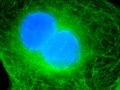
 Binucleated cell.jpg 514 × 386; 45 KB
-
 Cell divisions in Arabidopsis primary root meristem cells.tif 1,527 × 513; 765 KB
-
 Chromatin bridge stained with DAPI 2.tiff 1,392 × 1,040; 4.14 MB
-
 Cortical and mitotic microtubules in Arabidopsis primary root meristem cells.tif 1,525 × 504; 1,003 KB
-
 Cutie in mitosis.jpg 1,004 × 1,002; 835 KB
-
 Different mitotic stages in Arabidopsis primary root meristem cells.tif 774 × 256; 287 KB
-
 Dividing Cell Fluorescence-ru.jpg 554 × 554; 165 KB
-
 Dividing Cell Fluorescence-uk.jpg 554 × 554; 184 KB
-
 Dividing Cell Fluorescence.jpg 554 × 554; 58 KB
-
-
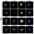
 Endogenous hMad1.png 575 × 873; 511 KB
-

-

-
 MAX MI DAPI 9-07-2015 A2 well.png 9,136 × 9,116; 62.29 MB
-
 Metaphase anaphase.png 232 × 153; 28 KB
-
 Mitosepanel es.jpg 1,050 × 246; 27 KB
-
 Mitosepanel-rus.tif 941 × 234; 234 KB
-
 Mitosepanel.jpg 1,050 × 246; 39 KB
-

-
 Mitotic spindle in Arabidopsis primary root meristem cells.tif 615 × 201; 195 KB
-
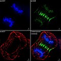
 SiCENP-E metaphase.png 501 × 510; 97 KB
-
 Spindle with Four Poles (8744518858).jpg 448 × 446; 93 KB
-
 The Biological bulletin (20353224406).jpg 1,806 × 1,582; 1,022 KB


_and_chromosomes_(red-yellow).png)


.jpg)
.jpg)