Category:Notochord
Jump to navigation
Jump to search
flexible rod-shaped body found in embryos of all chordates | |||||
| Upload media | |||||
| Instance of |
| ||||
|---|---|---|---|---|---|
| Subclass of |
| ||||
| Part of |
| ||||
| |||||
Media in category "Notochord"
The following 82 files are in this category, out of 82 total.
-
-

-
 Amphibia presumptive organ-forming areas in the late blastula and beginning gastrula.jpg 1,164 × 1,581; 1.61 MB
-
 Amphioxus Stage in which the external apertures of the body.jpg 616 × 2,235; 390 KB
-

-

-

-
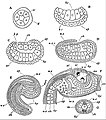
 Amphioxus Transverse sections through embryos of different ages.jpg 1,741 × 1,206; 1.09 MB
-
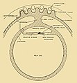
 Aves Diagrammatic transverse section of a chick embryo.jpg 1,057 × 862; 471 KB
-
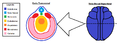
 Aves FGF signalling in mesoderm migration.jpg 2,152 × 612; 371 KB
-
-

-
-
-

-
-
-
 Aves View of the dorsal surface of a thirty-six-hour chick embryo.jpg 1,086 × 1,604; 918 KB
-
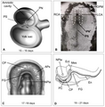
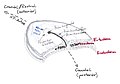
 Branchiostoma formation of the neural tube, notochord, mesoderm, and coelom.jpg 1,172 × 857; 562 KB
-
 Caudata process of invagination and mesoderm-formation.jpg 805 × 823; 698 KB
-

-
-

-
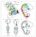
 Diagrams and images of human embryos at the gastrula stage.png 3,128 × 3,193; 804 KB
-
-
 Diversity-13-00462-g001.png 3,397 × 2,422; 1.39 MB
-
 Diversity-13-00462-g002.png 3,066 × 2,142; 1.43 MB
-
 EB1911 Tunicata - Stages in the Embryology of a Simple Ascidian.jpg 805 × 913; 216 KB
-
 Embryology (1949) (21285693065).jpg 1,035 × 1,115; 581 KB
-
 Embryonic myogenesis mouse.jpg 1,261 × 1,278; 938 KB
-
-
 Esquema corte transversal notocorda.png 535 × 291; 28 KB
-
 Formation and patterning of the mouse neural tube.png 1,305 × 1,505; 636 KB
-
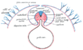
 Formation of notochord by primitive streak.jpg 1,091 × 1,071; 167 KB
-
 Fundulus heteroclitus presumptive organ-forming areas of the blastoderm.jpg 1,020 × 796; 779 KB
-

-
 Gray29.png 500 × 306; 15 KB
-
 Gray898.png 727 × 539; 297 KB
-
 Gray982.png 374 × 700; 25 KB
-
 Lungs of Protopterus dolloi.JPG 1,200 × 643; 568 KB
-
 Neurula.png 873 × 317; 21 KB
-
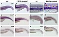 Not all tissues receiving notochord-emitted signals are affected by U0126 treatment.jpg 2,362 × 1,496; 1.48 MB
-
 Origin of Vertebrates Fig 163.png 1,425 × 451; 85 KB
-
 P270b The captain of the Omul eating the raw spinal cord of the sturgeon.jpg 1,251 × 1,573; 688 KB
-
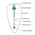
 Primitiv Node.jpg 720 × 504; 42 KB
-
 Protopterus dolloi Pulmo Hepar Chorda.png 1,200 × 643; 1.69 MB
-
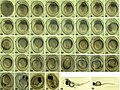 Pseudorasbora parva (10.3897-zoologia.35.e22162) Figures 2–39.jpg 1,997 × 1,494; 1.42 MB
-
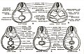
 Selachimorpha presumptive organ-forming areas in the blastoderm.jpg 981 × 801; 617 KB
-
-
-
-
Structural-Requirements-for-PACSINSyndapin-Operation-during-Zebrafish-Embryonic-Notochord-pone.0008150.s010.ogv 6.2 s, 1,280 × 720; 1.96 MB
-
-
 The biology of the frog (1927) (19759918144).jpg 2,042 × 1,892; 1.03 MB
-
 The Farm-poultry (1901) (14596885780).jpg 1,310 × 2,112; 408 KB
-
 The primitive streak and notochordal canal in Chelonia (Plate I) BHL4780730.jpg 2,577 × 1,849; 585 KB
-
 The primitive streak and notochordal canal in Chelonia (Plate II) BHL4780734.jpg 2,599 × 1,885; 3.31 MB
-
 The primitive streak and notochordal canal in Chelonia (Plate III) BHL4780738.jpg 2,104 × 1,877; 392 KB
-
 The primitive streak and notochordal canal in Chelonia (Plate V) BHL4780746.jpg 2,174 × 3,571; 849 KB
-
 The primitive streak and notochordal canal in Chelonia (Plate VI) BHL4780750.jpg 2,126 × 3,571; 1.01 MB
-
 The primitive streak and notochordal canal in Chelonia (Plate VII) BHL4780754.jpg 2,118 × 3,526; 778 KB
-
 The primitive streak and notochordal canal in Chelonia (Plate VIII) BHL4780758.jpg 2,555 × 1,863; 583 KB
-
 The primitive streak and notochordal canal in Chelonia (Plate X) BHL4780766.jpg 2,118 × 3,526; 1.49 MB
-
 The primitive streak and notochordal canal in Chelonia (Plate XI) BHL4780770.jpg 2,118 × 3,526; 1.13 MB
-
 The zebrafish Pacsin3 orthologue.jpg 714 × 995; 523 KB
-
-
-
-
-
-
-
-
-
-
-

-

-

-
 Zebrafish Early midline defects in pacsin3 morphants.jpg 690 × 966; 986 KB
-

-
 Zebrafish Embryonic Notochord Development The pacsin3 MO phenotype.jpg 843 × 895; 831 KB
-
 Zebrafish Structural requirements for Syndapin operation.jpg 1,091 × 717; 525 KB


_(20047812224).jpg)
_(20661197932).jpg)
,_myotome_(muscle-producer),_and_sclerotome_(skeleton-producer).jpg)


_(21285693065).jpg)


_Figures_2%E2%80%9339.jpg)

_analysis_of_the_posterior_tissue_precursors_during_posterior_axis_formation_chicken_embryo.jpg)
_(19759918144).jpg)
_(14596885780).jpg)
_BHL4780730.jpg)
_BHL4780734.jpg)
_BHL4780738.jpg)
_BHL4780746.jpg)
_BHL4780750.jpg)
_BHL4780754.jpg)
_BHL4780758.jpg)
_BHL4780766.jpg)
_BHL4780770.jpg)


