Category:Human radius
Jump to navigation
Jump to search
 Human anatomy Radius
| ||
|---|---|---|
Subcategories
This category has the following 10 subcategories, out of 10 total.
3
- 3D data of human radius (2 F)
A
- Animations of human radius (4 F)
F
H
- Humeroradial joints (15 F)
O
- Ossification of human radius (3 F)
P
- Photographs of human radius (16 F)
V
- Videos of human radius (4 F)
Media in category "Human radius"
The following 104 files are in this category, out of 104 total.
-
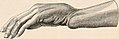 "Dinner fork" Deformity.jpg 1,604 × 526; 232 KB
-
 American quarterly of roentgenology (1912) (14570600039).jpg 776 × 1,632; 581 KB
-
 Anterior View of Right Distal Radius.png 1,152 × 2,048; 1.63 MB
-
 Anterior View of Right Proximal Radius.png 1,152 × 2,048; 1.36 MB
-
 Bidloo Ontleding 1690 71.jpg 1,200 × 1,614; 322 KB
-
 Buckle fracture of the Radius.svg 512 × 622; 1.04 MB
-
 Cunningham’s Text-book of Anatomy (1914) - Fig 208.png 1,038 × 1,268; 954 KB
-
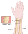
 Displaced distal radius fracture.jpg 1,494 × 1,513; 433 KB
-
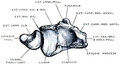
 Dixon's Manual of human osteology (1912) - Fig 041.png 981 × 2,031; 988 KB
-
 Dixon's Manual of human osteology (1912) - Fig 042.png 669 × 2,031; 577 KB
-
 Dixon's Manual of human osteology (1912) - Fig 043.png 780 × 1,983; 408 KB
-
 Dixon's Manual of human osteology (1912) - Fig 044.png 1,143 × 2,052; 866 KB
-
 Dixon's Manual of human osteology (1912) - Fig 045.png 1,073 × 542; 301 KB
-
 Dixon's Manual of human osteology (1912) - Fig 046.png 1,203 × 567; 289 KB
-
 Dixon's Manual of human osteology (1912) - Fig 047.png 951 × 1,983; 463 KB
-
 Drawing RADIUS 1812.svg 389 × 174; 7 KB
-
 Forearm cut.png 550 × 422; 104 KB
-
 Full Anterior View of Right Radius.png 1,152 × 2,048; 477 KB
-
 Full Lateral View of Right Radius.png 1,152 × 2,048; 402 KB
-
 Full Medial View of Right Radius.png 1,152 × 2,048; 420 KB
-
 Full Posterior View of Right Radius.png 1,152 × 2,048; 441 KB
-
 Gerrish's Text-book of Anatomy (1902) - Fig. 168.png 740 × 1,767; 625 KB
-
 Gerrish's Text-book of Anatomy (1902) - Fig. 169.png 786 × 1,761; 619 KB
-
 Gerrish's Text-book of Anatomy (1902) - Fig. 170.png 786 × 1,803; 614 KB
-
 Gerrish's Text-book of Anatomy (1902) - Fig. 171.png 765 × 1,803; 464 KB
-
 Gray217.png 251 × 400; 9 KB
-
 Gray218.png 170 × 450; 10 KB
-
 Gray420-ar.png 329 × 550; 81 KB
-
 Gray420.png 329 × 550; 34 KB
-
 Gray421.png 550 × 312; 26 KB
-
 Holden's human osteology (1899) - Plt52.png 1,683 × 2,734; 2.38 MB
-
 Holden's human osteology (1899) - Plt53.png 1,704 × 2,812; 2.68 MB
-
 Human radius.stl 5,120 × 2,880; 22.37 MB
-
 Jud-naiwan.gif 430 × 430; 7 KB
-
 Jud-naiwan.svg 430 × 430; 69 KB
-
 Lateral View of Right Distal Radius.png 1,152 × 2,048; 923 KB
-
 Lateral View of Right Proximal Radius.png 1,152 × 2,048; 892 KB
-
 Left radius - close-up - animation.gif 450 × 450; 446 KB
-
 Left Radius.stl 5,120 × 2,880; 12.38 MB
-
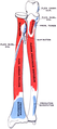
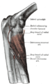
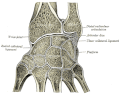
 Ligaments of the elbow.jpg 1,000 × 1,000; 154 KB
-
 Medial View of Right Distal Radius.png 1,152 × 2,048; 1.34 MB
-
 Medial View of Right Proximal Radius.png 1,152 × 2,048; 1.46 MB
-
 Medical X-Ray imaging OCU06 nevit.jpg 1,784 × 2,384; 463 KB
-
 Medical X-Ray imaging TJV07 nevit.jpg 1,974 × 2,486; 855 KB
-
 Medical X-Ray imaging VNH07 nevit.jpg 1,974 × 2,486; 704 KB
-
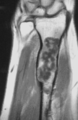 Morbus Ollier Radius MRT T1.png 400 × 615; 92 KB
-
 Morbus Ollier Radius Roentgen.png 1,341 × 1,720; 963 KB
-
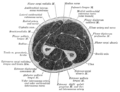
 Muscles of upper limb.(cross section - human cadaver)-zh.jpg 960 × 720; 185 KB
-
 Ospoignet.gif 550 × 430; 56 KB
-
 Posterior Radius Proximal.png 1,920 × 1,080; 271 KB
-
 Posterior View of Right Distal Radius.png 1,152 × 2,048; 1.04 MB
-
 Posterior View of Right Proximal Radius.png 1,152 × 2,048; 931 KB
-
 Quain's elements of anatomy (1891) - Vol2 Part1- Fig 094.png 895 × 2,796; 1.37 MB
-
 Quain's elements of anatomy (1891) - Vol2 Part1- Fig 097.png 764 × 2,504; 846 KB
-
 Quain's elements of anatomy (1891) - Vol2 Part1- Fig 098.png 942 × 510; 329 KB
-
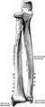
 RADIO E ULNA.jpg 1,061 × 1,886; 177 KB
-
 Radius - animation.gif 450 × 450; 2.33 MB
-
 Radius - animation2.gif 450 × 450; 3.09 MB
-
 Radius - anterior view.png 4,500 × 4,500; 3.63 MB
-
 Radius - anterior view2.png 4,500 × 4,500; 4.51 MB
-
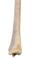
 Radius - anterior.jpg 4,108 × 1,676; 2.7 MB
-
 Radius - detail of bone tissue (distal end).jpg 3,320 × 2,840; 3.73 MB
-
 Radius - distal end.jpg 3,280 × 2,160; 2.92 MB
-
 Radius - lateral view.png 4,500 × 4,500; 2.29 MB
-
 Radius - lateral view2.png 4,500 × 4,500; 3.03 MB
-
 Radius - posterior 2.jpg 4,608 × 3,456; 4.65 MB
-
 Radius - posterior view.png 4,500 × 4,500; 3.63 MB
-
 Radius - posterior view2.png 4,500 × 4,500; 5.77 MB
-
 Radius - posterior.jpg 4,320 × 1,752; 2.7 MB
-
 Radius - proximal end.jpg 2,976 × 2,301; 2.5 MB
-
 Radius 10deg lateral break gnangarra.jpg 618 × 1,535; 155 KB
-
Radius and ulna (preview) - Human Anatomy Kenhub 1.webm 2 min 3 s, 1,280 × 720; 79.13 MB
-
 Radius ant pos num.JPG 748 × 1,627; 169 KB
-
 Radius ant.jpg 348 × 1,627; 127 KB
-
 Radius Anteiror Distal 01.png 1,920 × 1,080; 294 KB
-
 Radius Anteiror Proximal.png 1,920 × 1,080; 276 KB
-
Radius Bone Anatomy by Jason Christian.webm 40 s, 1,280 × 720; 8.29 MB
-
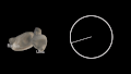 Radius Bone and Radius of a circle comparison.gif 800 × 450; 1.7 MB
-
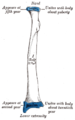
 Radius Bone with Labels.gif 800 × 800; 2.85 MB
-
 Radius post.jpg 348 × 1,627; 125 KB
-
 Radius Posterior Distal.png 1,920 × 1,080; 334 KB
-
Radius Walk-thru by Bob Myers.webm 58 s, 640 × 480; 1.17 MB
-
 Radius-epiphysiolyse01.jpg 366 × 619; 41 KB
-
 Radius.JPG 512 × 1,695; 94 KB
-
 Radius.jpg 960 × 720; 61 KB
-
 Radius2.jpg 960 × 720; 48 KB
-
 Radius3.jpg 960 × 720; 42 KB
-
 Radius4.jpg 960 × 720; 49 KB
-
-
 Right hand-3d Arabic YM.jpg 522 × 600; 90 KB
-
 RightHumanAnteriorDistalRadiusUlnaCarpals.jpg 2,661 × 1,681; 687 KB
-
 Rx-human ulna radio.jpg 2,549 × 3,508; 2.02 MB
-
 Slide3TTTTT.JPG 960 × 720; 86 KB
-
 Sobo 1909 120.png 1,520 × 604; 2.63 MB
-
 Sobo 1909 121.png 684 × 2,376; 4.66 MB
-
 Sobo 1909 122.png 884 × 2,312; 5.86 MB
-
 Sobo 1909 123.png 928 × 2,548; 6.78 MB
-
 Sobo 1909 202.png 2,308 × 1,288; 8.52 MB
-
 Supinator.png 255 × 550; 31 KB
-
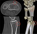 Tuberculum dorsale radii 21W - CT - 001 - Annotation.jpg 603 × 569; 68 KB
-
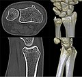 Tuberculum dorsale radii 21W - CT - 001.jpg 603 × 569; 51 KB
-
 Tumor Myeloma Radius.JPG 759 × 849; 30 KB
-
 Unterarmknochen ap.png 1,559 × 492; 509 KB
-
_(14570600039).jpg)


_-_Fig_208.png)

_-_Fig_041.png)
_-_Fig_044.png)
_-_Fig_045.png)
_-_Fig_046.png)
_-_Fig_047.png)


_-_Fig._168.png)
_-_Fig._169.png)
_-_Fig._170.png)
_-_Fig._171.png)


_-_Plt52.png)
_-_Plt53.png)


-zh.jpg)


_-_Vol2_Part1-_Fig_098.png)


.jpg)


_(14741652866).jpg)
_-_Fig_042.png)
_-_Fig_043.png)
_-_Vol2_Part1-_Fig_094.png)
_-_Vol2_Part1-_Fig_097.png)