Category:Gastrulation
Jump to navigation
Jump to search
Phase in the early embryonic development of most animals.  | |||||
| Upload media | |||||
| Instance of | |||||
|---|---|---|---|---|---|
| Subclass of |
| ||||
| Has part(s) |
| ||||
| Follows |
| ||||
| |||||
Subcategories
This category has the following 7 subcategories, out of 7 total.
Media in category "Gastrulation"
The following 126 files are in this category, out of 126 total.
-

-

-

-

-
 ADMP restricts the size of the organizer domain Spemann's organizer.jpg 818 × 665; 397 KB
-
 ALK1 is required to restrict the size of the organizer domain Spemann's organizer.jpg 1,374 × 1,026; 1.27 MB
-

-

-
 Antedon rosacea early stages.jpg 801 × 735; 425 KB
-
 Arquénteron.jpg 1,066 × 1,061; 73 KB
-
 Avian development before gastrulation.jpg 2,767 × 3,354; 1.41 MB
-

-
 Branchiostoma formation of the neural tube, notochord, mesoderm, and coelom.jpg 1,172 × 857; 562 KB
-
 Caudata process of invagination and mesoderm-formation.jpg 805 × 823; 698 KB
-

-

-

-
-
 CENTROLECITHES.png 351 × 540; 12 KB
-

-
Control-of-Directed-Cell-Migration-In-Vivo-by-Membrane-to-Cortex-Attachment-pbio.1000544.s009.ogv 6.2 s, 264 × 264; 701 KB
-
Control-of-Directed-Cell-Migration-In-Vivo-by-Membrane-to-Cortex-Attachment-pbio.1000544.s010.ogv 6.2 s, 264 × 264; 388 KB
-
Control-of-Directed-Cell-Migration-In-Vivo-by-Membrane-to-Cortex-Attachment-pbio.1000544.s011.ogv 6.2 s, 264 × 264; 454 KB
-
Control-of-Directed-Cell-Migration-In-Vivo-by-Membrane-to-Cortex-Attachment-pbio.1000544.s012.ogv 7.6 s, 418 × 416; 1.51 MB
-
Control-of-Directed-Cell-Migration-In-Vivo-by-Membrane-to-Cortex-Attachment-pbio.1000544.s013.ogv 7.5 s, 419 × 416; 680 KB
-
Control-of-Directed-Cell-Migration-In-Vivo-by-Membrane-to-Cortex-Attachment-pbio.1000544.s014.ogv 6.5 s, 512 × 512; 532 KB
-
-
Control-of-Directed-Cell-Migration-In-Vivo-by-Membrane-to-Cortex-Attachment-pbio.1000544.s016.ogv 11 s, 1,024 × 529; 2.41 MB
-
Control-of-Directed-Cell-Migration-In-Vivo-by-Membrane-to-Cortex-Attachment-pbio.1000544.s017.ogv 5.7 s, 370 × 383; 589 KB
-
Control-of-Directed-Cell-Migration-In-Vivo-by-Membrane-to-Cortex-Attachment-pbio.1000544.s018.ogv 6.5 s, 370 × 370; 738 KB
-
-

-

-
Deficient-Induction-Response-in-a-Xenopus-Nucleocytoplasmic-Hybrid-pbio.1001197.s005.ogv 13 s, 434 × 668; 351 KB
-
 DELAMINATION.png 934 × 573; 26 KB
-

-

-
-
 Differentiation of the mesoderm in holoblastic and meroblastic types of development.jpg 1,617 × 1,467; 1,022 KB
-
 Diversity of vertebrate gastrulation.jpg 1,073 × 1,021; 415 KB
-
 Early gastrulation in amphibian embryos.png 3,570 × 1,358; 1,007 KB
-

-
 EMBRIO 1.jpg 571 × 116; 14 KB
-
 EMBRIO 2.jpg 943 × 729; 59 KB
-
 EMBRIO 3.jpg 341 × 346; 17 KB
-
 EMBRIO 4.jpg 376 × 193; 10 KB
-
 EMBRIO 5.jpg 398 × 283; 10 KB
-
 EPIBOLIE.png 934 × 573; 26 KB
-
 Epiboly - Advanced stage.jpg 1,080 × 1,920; 252 KB
-
 Epiboly Drawing.png 2,550 × 2,073; 5.64 MB
-
 Example of a Gastruloid.png 673 × 336; 134 KB
-
 Experimental manipulation of the gastrulation mode in different organisms.jpg 1,166 × 1,480; 850 KB
-
 Fig 3. Proceso de gastrulación en el nemátodo C.elegans..png 1,278 × 427; 121 KB
-
 Formation of the primitive body plan following gastrulation in the mouse.png 1,279 × 1,187; 1,016 KB
-
 Frog egg gray crescent.jpg 1,060 × 816; 133 KB
-
 Gastrulatie.png 1,780 × 1,520; 1.6 MB
-
 Gastrulation (01).jpg 1,001 × 980; 289 KB
-
 Gastrulation forms in vertebrates.jpeg 1,280 × 1,173; 88 KB
-

-
 Gastrulation of Asterina pectinifer.jpg 1,351 × 521; 212 KB
-
 Gastrulatsiooni toimumine.jpg 690 × 354; 67 KB
-
 Gastrulação.png 695 × 349; 13 KB
-
 HETEROLECITHES.png 322 × 224; 5 KB
-

-

-

-

-
 INGRESSION.png 934 × 573; 26 KB
-
 Koller's Sickle and Avian Gastrulation.jpg 1,967 × 1,968; 863 KB
-
 Koller's sickle and its role in avian gastrulation.jpg 271 × 448; 47 KB
-
 Kudede päritolu gastrulatsioonis.jpg 600 × 700; 74 KB
-

-
 Left-right asymmetry in the sea urchin - journal.pbio.1001404.g001.png 1,459 × 420; 235 KB
-

-
 Mesoderm-ring to -crescent transition.jpg 1,173 × 1,826; 1.01 MB
-
 Meyers b5 s0350 b1.png 570 × 218; 53 KB
-
Modeling-Gastrulation-in-the-Chick-Embryo-Formation-of-the-Primitive-Streak-pone.0010571.s002.ogv 17 s, 648 × 492; 16.95 MB
-
Modeling-Gastrulation-in-the-Chick-Embryo-Formation-of-the-Primitive-Streak-pone.0010571.s003.ogv 9.5 s, 700 × 350; 5.23 MB
-
Modeling-Gastrulation-in-the-Chick-Embryo-Formation-of-the-Primitive-Streak-pone.0010571.s004.ogv 21 s, 700 × 350; 17.02 MB
-
Modeling-Gastrulation-in-the-Chick-Embryo-Formation-of-the-Primitive-Streak-pone.0010571.s005.ogv 22 s, 1,050 × 350; 19.89 MB
-
Modeling-Gastrulation-in-the-Chick-Embryo-Formation-of-the-Primitive-Streak-pone.0010571.s007.ogv 15 s, 700 × 700; 10.8 MB
-
Modeling-Gastrulation-in-the-Chick-Embryo-Formation-of-the-Primitive-Streak-pone.0010571.s008.ogv 30 s, 1,050 × 350; 22.29 MB
-
Modeling-Gastrulation-in-the-Chick-Embryo-Formation-of-the-Primitive-Streak-pone.0010571.s009.ogv 0.0 s, 1,056 × 352; 22.13 MB
-
 Morphological differences between human and mouse gastrulation.jpg 2,994 × 3,411; 397 KB
-
 Nematode Variations in gastrulation.jpg 1,800 × 1,909; 1.74 MB
-
 Neurula.png 873 × 317; 21 KB
-
 OLIGOLECITHES.png 324 × 235; 4 KB
-

-
 Paracentrotus lividus gastrula.jpg 1,245 × 934; 1.08 MB
-
PhOTO-Zebrafish-A-Transgenic-Resource-for-In-Vivo-Lineage-Tracing-during-Development-and-pone.0032888.s005.ogv 1.7 s, 1,984 × 1,952; 2.06 MB
-
PhOTO-Zebrafish-A-Transgenic-Resource-for-In-Vivo-Lineage-Tracing-during-Development-and-pone.0032888.s006.ogv 1.7 s, 1,984 × 1,476; 1.5 MB
-
PhOTO-Zebrafish-A-Transgenic-Resource-for-In-Vivo-Lineage-Tracing-during-Development-and-pone.0032888.s007.ogv 2.5 s, 1,972 × 1,972; 5.84 MB
-
PhOTO-Zebrafish-A-Transgenic-Resource-for-In-Vivo-Lineage-Tracing-during-Development-and-pone.0032888.s008.ogv 2.5 s, 1,972 × 1,476; 3.08 MB
-
-

-
Regulation-of-Embryonic-Cell-Adhesion-by-the-Prion-Protein-pbio.1000055.sv001.ogv 8.0 s, 299 × 299; 1.19 MB
-
Regulation-of-Embryonic-Cell-Adhesion-by-the-Prion-Protein-pbio.1000055.sv002.ogv 8.0 s, 356 × 357; 1.66 MB
-
Regulation-of-Embryonic-Cell-Adhesion-by-the-Prion-Protein-pbio.1000055.sv003.ogv 12 s, 158 × 155; 88 KB
-
Regulation-of-Embryonic-Cell-Adhesion-by-the-Prion-Protein-pbio.1000055.sv004.ogv 12 s, 158 × 156; 92 KB
-
Regulation-of-Embryonic-Cell-Adhesion-by-the-Prion-Protein-pbio.1000055.sv005.ogv 2.1 s, 1,024 × 1,024; 1.68 MB
-
Regulation-of-Embryonic-Cell-Adhesion-by-the-Prion-Protein-pbio.1000055.sv006.ogv 12 s, 523 × 512; 340 KB
-
 RG347357480 Fig1.png 850 × 377; 60 KB
-
 Rho kinase (ROCK1) is expressed during the mammalian gastrulation.jpg 795 × 563; 466 KB
-
 Sea Urchin Gastrulation.png 752 × 300; 187 KB
-
-
 Staging Human Gastrulation.jpg 3,493 × 3,010; 538 KB
-
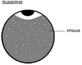 TELOLECITHES.png 487 × 384; 5 KB
-

-
 The Biological bulletin (19758011533).jpg 2,004 × 1,310; 608 KB
-
 The evolution of man (1920) plate III.jpg 753 × 1,173; 1.21 MB
-
-
-
-
-
-
-
-
-
-
-
-
 Tracking of EB3-GFP movement shows microtubule growth in Xenopus explants.png 2,243 × 2,960; 1.42 MB
-
-
Xenopus-Nucleocytoplasmic-Hybrid.ogv 15 s, 292 × 782; 557 KB
-
-


_(20047812224).jpg)


,_myotome_(muscle-producer),_and_sclerotome_(skeleton-producer).jpg)


.jpg)


_is_expressed_during_the_mammalian_gastrulation.jpg)
_analysis_of_the_posterior_tissue_precursors_during_posterior_axis_formation_chicken_embryo.jpg)


.jpg)
_plate_III.jpg)
_(14760848311).jpg)
_and_CD31_(green)_at_different_time_points_(24_week)_after_vascular_transplantation..png)