Category:Fish anatomy illustrations
Jump to navigation
Jump to search
Subcategories
This category has the following 3 subcategories, out of 3 total.
Media in category "Fish anatomy illustrations"
The following 158 files are in this category, out of 158 total.
-
 A. Monro, "The structure...of fishes..." Wellcome L0028245.jpg 1,198 × 1,676; 898 KB
-
 Acanthodes skull.jpg 1,280 × 1,054; 93 KB
-
 Acta Societatis Scientiarum Fennicae (1909) (16151586323).jpg 3,300 × 2,162; 1.29 MB
-
 Acta Societatis Scientiarum Fennicae (1909) (16564255797).jpg 3,358 × 2,130; 1.39 MB
-
 Acta Societatis Scientiarum Fennicae (1909) (16583955698).jpg 3,216 × 2,160; 1.33 MB
-
 Acta Societatis Scientiarum Fennicae (1909) (16745646696).jpg 3,402 × 2,180; 1.37 MB
-
 Acta Societatis Scientiarum Fennicae (1909) (16771520285).jpg 3,312 × 2,148; 1.41 MB
-

-

-
-
-
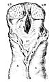
 Anoxypristis cuspidata rosrrum.jpg 663 × 111; 24 KB
-
 Archives du Muséum d'Histoire Naturelle, Paris BHL25099466.jpg 2,273 × 3,198; 719 KB
-
 Archives du Muséum d'Histoire Naturelle, Paris BHL25099470.jpg 2,273 × 3,198; 562 KB
-
 Archives du Muséum d'Histoire Naturelle, Paris BHL25099472.jpg 2,273 × 3,198; 609 KB
-
 Bachforelle osmoregulatoin bw en2.png 1,000 × 500; 170 KB
-
 Balık.png 520 × 364; 63 KB
-
 Britannica Shark Blue Shark Teeth.png 925 × 939; 30 KB
-
 Britannica Shark Scyllium canicula Teeth.png 493 × 323; 9 KB
-
 Britannica Shark Tope Teeth.png 165 × 308; 4 KB
-
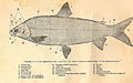
 British and Irish Salmonidæ (Plate I) (8539692922).jpg 1,175 × 1,840; 250 KB
-
 British and Irish Salmonidæ (Plate II) (8539693202).jpg 1,843 × 1,175; 260 KB
-
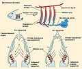
 Brockhaus-Efron Electric Organs 1.jpg 522 × 699; 81 KB
-
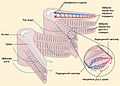
 Brockhaus-Efron Electric Organs 2.jpg 870 × 625; 169 KB
-

-
 Bulletin de la Société impériale des naturalistes de Moscou (1829-1917.) (20248818319).jpg 3,064 × 2,280; 1.06 MB
-
 Bulletin du Muséum d'histoire naturelle (1917) (20251891830).jpg 3,760 × 2,518; 2.19 MB
-
 California fish and game (20324755990).jpg 3,232 × 1,960; 476 KB
-
 California fish and game (20325147850).jpg 1,366 × 1,890; 314 KB
-
 California fish and game (20487417686).jpg 1,970 × 3,270; 442 KB
-
 Cod-icelandic.svg 577 × 237; 23 KB
-

-
 Cross section of a standard fish.jpg 933 × 925; 177 KB
-
 Cross section of standard fish.svg 512 × 492; 50 KB
-
 Cuvier-99-Malaptérure.jpg 1,561 × 1,134; 958 KB
-
 Dasyatis say njsm (annotated).jpg 737 × 445; 152 KB
-
-
 Eastman1901-web.jpg 490 × 150; 22 KB
-
 EB1911 Ichthyology - Chondrocranium of a young Lepidosiren.jpg 859 × 502; 91 KB
-
 EB1911 Ichthyology - condition of the Conus.jpg 444 × 355; 48 KB
-

-
 EB1911 Ichthyology - Cranial nerves of a Fish.jpg 848 × 394; 99 KB
-
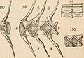
 EB1911 Ichthyology - Morphology of the vertebral column.jpg 877 × 877; 266 KB
-
 EB1911 Ichthyology - Venous System of Polypterus (larva).jpg 412 × 690; 65 KB
-
 EB1911 Ichthyology - Venous System of Protopterus.jpg 392 × 1,030; 90 KB
-
 Elementary zoology (1902) (21241557581).jpg 2,704 × 2,852; 2.59 MB
-
 Encéphale du congre commun.jpg 285 × 559; 72 KB
-
 Evolution ear fishes.jpg 750 × 525; 59 KB
-
 Field book of ponds and streams (Page 325) BHL4950049.jpg 1,835 × 3,296; 593 KB
-
 Figure 39 01 04.jpg 994 × 1,033; 436 KB
-
 Fish anatomy (berycid).png 520 × 364; 72 KB
-
 Fish Brain.jpg 906 × 533; 153 KB
-
 Fish fin anatomy Salmonidae.svg 661 × 239; 6.15 MB
-
 Fish gill structure.jpg 912 × 656; 239 KB
-
 Fish kidney.jpg 931 × 775; 138 KB
-
 Fish length.jpg 343 × 145; 10 KB
-
 Fish mouths.orizz.png 703 × 164; 15 KB
-
 Fish-anatomy.svg 1,038 × 541; 21 KB
-
 Fishes (1907) (14777170862).jpg 2,896 × 1,996; 419 KB
-
 FishGills1.jpg 911 × 756; 233 KB
-
 FishGills2.jpg 912 × 656; 206 KB
-
-
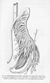
 FMIB 38604 Dorsal Spine of Siluridae.jpeg 707 × 1,219; 81 KB
-
 FMIB 39114 View of normal floor of mouth of adult brook trout.jpeg 266 × 379; 14 KB
-
 FMIB 46089 Thornback Ray.jpeg 905 × 1,277; 365 KB
-
 FMIB 46204 Banks' Oarfish - Caudal Extremity.jpeg 815 × 567; 102 KB
-
-
 FMIB 47050 Dentition of the Blue Shark (Carcharias glaucus).jpg 635 × 663; 84 KB
-
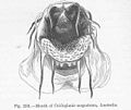 FMIB 47146 Mouth of Cnidoglanis megastoma, Australia.jpeg 643 × 539; 82 KB
-

-
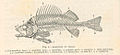 FMIB 47969 Skeleton of Perch.jpeg 1,037 × 477; 106 KB
-

-
 FMIB 47973 Internal Anatomy of Carp.jpeg 1,127 × 463; 137 KB
-
 FMIB 47974 Internal Anatomy of Carp.jpeg 1,078 × 373; 102 KB
-
 FMIB 48114 Head of Salmo salar.jpeg 546 × 359; 52 KB
-
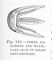
 FMIB 48116 Vomer of Salmo obtusirostris.jpeg 201 × 277; 11 KB
-
-
 FMIB 48123 Front view of Vomer of Salmo marsiglii.jpeg 283 × 194; 11 KB
-
 FMIB 50416 Anatomy of the Carp.jpeg 658 × 1,061; 188 KB
-
 FMIB 50417 Fishes' Eye.jpeg 482 × 368; 26 KB
-
 FMIB 51375 Gill of Mehnaden.jpeg 1,068 × 512; 198 KB
-
 FMIB 51552 Dissection of the Blue-green sunfish, Apomotis cyanellus Rafinesque.jpeg 1,833 × 1,038; 475 KB
-

-

-

-

-

-
 FMIB 51664 Teeth of the Heptranchias indicus Gmelin.jpg 695 × 557; 71 KB
-
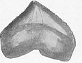 FMIB 51679 Teeth of Corax pristodontus.jpeg 458 × 352; 45 KB
-
 FMIB 51683 Brain of Monkfish, Squatina squatina L.jpeg 261 × 639; 36 KB
-
 FMIB 51826 Weberian apparatus and air-bladder of Carp.jpeg 831 × 404; 119 KB
-

-
-
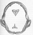
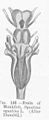
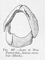 FMIB 52069 Jaws of Blue Parrot-fish, Scarus caeruleus (Bloch).jpeg 343 × 458; 46 KB
-

-
 FMIB 52343 Lateral view of Perugia xanthus.jpeg 513 × 330; 75 KB
-
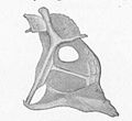
 FMIB 52354 Dorsal Plate of Pimelodus clarias.jpeg 269 × 418; 25 KB
-

-
 Gigantactis photo.JPG 1,795 × 1,488; 267 KB
-
 Hisonotus acuen-442-105-g004 (2).jpg 771 × 564; 75 KB
-
 Histoire des poissons BHL6231147.jpg 3,546 × 1,877; 679 KB
-
 Histoire des poissons BHL6231149.jpg 2,168 × 1,790; 276 KB
-
 Histoire naturelle des poissons (10438645606).jpg 3,200 × 1,928; 672 KB
-
 Histoire naturelle des poissons (10438648826).jpg 1,928 × 3,200; 648 KB
-
 Histoire naturelle des poissons (Pl. 138) (7950032790).jpg 1,893 × 3,200; 485 KB
-
 Histoire naturelle des poissons (Pl. 139) (7950033758).jpg 1,893 × 3,200; 583 KB
-
 Hunter Electric Eel 1775 cross-section.jpg 764 × 841; 112 KB
-
 Hunter Electric Eel Dissection 1775.jpg 1,053 × 651; 181 KB
-
 Internal organs of a fish.jpg 650 × 325; 53 KB
-
 Internal salmon anatomy.jpg 553 × 252; 43 KB
-
 Knollenorgan by Viktor Franz 1921.gif 348 × 292; 4 KB
-
 Loricaria-eye.png 749 × 331; 32 KB
-

-

-

-
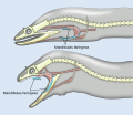 MandFar.svg 1,500 × 1,300; 524 KB
-
 Mauthner Cell axon cap schematic.svg 325 × 295; 23 KB
-
 Meyer Zeit-Vertreib 2 Tafel 052.jpg 1,466 × 896; 180 KB
-
 Meyer Zeit-Vertreib 2 Tafel 092.jpg 1,456 × 897; 183 KB
-
 NIE 1905 Fin.jpg 322 × 232; 29 KB
-
 Zoologie. Poissons du Nil. Hétérobranches. Détails anatomiques (NYPL b14212718-1268516).jpg 4,091 × 5,297; 4.77 MB
-
 Ossicini di Weber.jpg 60 × 107; 3 KB
-
 Ossicini di Weber.png 60 × 107; 15 KB
-
 Our country's fishes and how to know them (Page 42, Fig. 23) BHL20965883.jpg 3,680 × 2,554; 1 MB
-
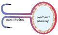 PecherzPlawny-grafik.svg 330 × 200; 58 KB
-
 Pira posẽ.jpg 2,376 × 884; 688 KB
-
 Pristis microdon LATHAM,1794.jpg 600 × 146; 15 KB
-
 Pristis pectinata rostrum.png 656 × 159; 163 KB
-
 PSM V04 D040 Head of a kelt.jpg 1,622 × 826; 260 KB
-
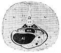 PSM V11 D012 Gar pike cross section.jpg 602 × 532; 73 KB
-
 PSM V11 D018 Gar pike longitudonal head section.jpg 1,301 × 398; 76 KB
-
-
 PSM V11 D545 Torpedo fish.jpg 1,059 × 1,438; 142 KB
-
 PSM V26 D354 Sword of young swordfish.jpg 1,052 × 2,168; 472 KB
-
 PSM V57 D064 Chologaster papilliferus with the tactile ridges.png 657 × 553; 99 KB
-
 PSM V57 D065 Lateral view of amblyopsis showing location of tactile ridges.png 1,820 × 1,077; 131 KB
-

-

-
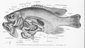
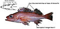 Pygmyrockfish.jpg 1,013 × 493; 42 KB
-
 PZSL1889Plate21a.png 1,856 × 2,910; 5.31 MB
-
 PZSL1889Plate21b.png 1,856 × 2,910; 5.18 MB
-

-
 Scientific investigations (1907) (14577457949).jpg 3,208 × 2,558; 834 KB
-
 Scientific investigations (1911) (14596235667).jpg 2,892 × 2,284; 1,000 KB
-
 Scientific investigations (1911) (14782741805).jpg 2,852 × 2,196; 1.02 MB
-
 Scientific investigations (1911) (14802596923).jpg 2,894 × 2,238; 1,010 KB
-
 Squatina squatina maxillary.jpg 811 × 1,000; 272 KB
-
 The Australian zoologist (1967) (20341900312).jpg 2,960 × 1,976; 3.09 MB
-
 The cranial anatomy of the mail-cheeked fishes (1909) (20520372028).jpg 2,970 × 1,620; 544 KB
-
 The fishes of Great Britain and Ireland (1880) (20566783560).jpg 1,508 × 874; 521 KB
-
 The Plagiostomia (Plate 50) (6001823198).jpg 1,242 × 1,726; 90 KB
-
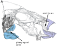 Toothed Oral and Pharyngeal Jaws (crop).tiff 1,482 × 1,223; 6.92 MB
-
 Toothed Oral and Pharyngeal Jaws.tif 2,260 × 2,408; 6.5 MB
-
 Transactions and proceedings of the New Zealand Institute (1887) (14773633611).jpg 1,588 × 2,888; 382 KB
-
-
 Trout fly-fishing in America (6308549575).jpg 3,114 × 2,033; 519 KB
-
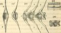
 Vestiges 11 fig 34 Cod vertebrae.jpg 1,824 × 1,331; 387 KB
-
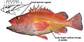 Yelloweyerockfish.jpg 1,013 × 513; 52 KB


_(16151586323).jpg)
_(16564255797).jpg)
_(16583955698).jpg)
_(16745646696).jpg)
_(16771520285).jpg)
_als_Clupea_alosa_1.Kiemenbogen_Fig_110a_(Matschie_et_al._1909).svg)
_(18143166321).jpg)
_(14762400394).jpg)
_(14779055565).jpg)


_(8539692922).jpg)
_(8539693202).jpg)


_(19812828294).jpg)
_(20248818319).jpg)
_(20251891830).jpg)
.jpg)
.jpg)
.jpg)
_(20670796735).jpg)


.jpg)
_(14776614074).jpg)


.jpg)
_(21241557581).jpg)


_BHL4950049.jpg)

.png)


_(14777170862).jpg)


.jpg)


_Family_Cyprinidon_Showing_nuptial_tubercles_and_intestines_coiled_about_the_air.jpeg)

_A_viviparous_fish_from_Lake_Patzcuaro,_Mexico_Family_Paciliidae.jpeg)


.jpeg)


.jpeg)

.jpg)


.jpg)
.jpg)
_(7950032790).jpg)
_(7950033758).jpg)


_(7038556209).jpg)
_(6005498597).jpg)
_(7038556901).jpg)


.jpg)


_BHL20965883.jpg)


.svg)
_(14577457949).jpg)
_(14596235667).jpg)
_(14782741805).jpg)
_(14802596923).jpg)

_(20341900312).jpg)
_(20520372028).jpg)
_(20566783560).jpg)
_(6001823198).jpg)
.jpg)


_(14773633611).jpg)