Category:Diseases and disorders of the eye and adnexa
Jump to navigation
Jump to search
Radiology: Ultrasound · Computed tomography · Magnetic resonance | Anatomical pathology: Gross pathology · Histopathology | Other: Fundus · Optical coherence tomography · Epidemiology (Map → World) | File format: SVG · Video recording | 

Categorization according to International Statistical Classification of Diseases and Related Health Problems, 10th Revision.[edit]
health condition negatively affecting the eye | |||||
| Upload media | |||||
| Instance of |
| ||||
|---|---|---|---|---|---|
| Subclass of |
| ||||
| |||||
Subcategories
This category has the following 54 subcategories, out of 54 total.
*
-
A
- Adie syndrome (1 F)
B
- Bitot's spots (2 F)
- Buphthalmos (2 F)
C
- Corectopia (1 F)
- Eye cysts (2 F)
D
F
G
- Globe rupture (3 F)
- Eye gumma (2 F)
H
I
K
- Keratic precipitate (2 F)
L
- Leukocoria (5 F)
N
O
P
S
V
W
- Waardenburg syndrome (18 F)
Pages in category "Diseases and disorders of the eye and adnexa"
This category contains only the following page.
Media in category "Diseases and disorders of the eye and adnexa"
The following 32 files are in this category, out of 32 total.
-
 120823-F-CF823-217 (7938218002).jpg 4,256 × 2,832; 6.01 MB
-
 2012-09-22 eye with disease.jpg 2,768 × 2,079; 958 KB
-
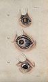 A sheet of three diagrams showing inflamed bloodshot eye def Wellcome V0015923EL.jpg 1,266 × 2,136; 1.53 MB
-
 A sheet of three diagrams showing inflamed bloodshot eye def Wellcome V0015923ER.jpg 1,233 × 2,086; 1.32 MB
-
 A sheet of three diagrams showing inflamed eye defects. Colo Wellcome V0015922EL.jpg 1,272 × 2,082; 1.71 MB
-
 A sheet of three diagrams showing inflamed eye defects. Colo Wellcome V0015922ER.jpg 1,251 × 2,088; 1.49 MB
-
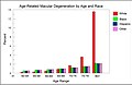 AMD by race and age from National Eye Institute data.jpg 1,882 × 1,216; 289 KB
-

-
 Bilateral222.jpg 666 × 247; 94 KB
-
 Eye discharge.jpg 1,690 × 1,044; 321 KB
-

-

-
-
 Irvine-Gass syndrome .png 1,920 × 1,329; 538 KB
-

-
 Macro Globe oculaire - Mélanome 55-o.apatho-52a-oeil.jpg 639 × 1,499; 500 KB
-
 Macro Globe oculaire - Mélanome 55-o.apatho-52p-oeil.jpg 735 × 540; 195 KB
-
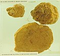 Macro Globe oculaire - Rétinoblastome 55-o.apatho-242a-oeil.jpg 1,676 × 1,547; 1.44 MB
-
 Macro Globe oculaire - Rétinoblastome 55-o.apatho-242p-oeil.jpg 1,563 × 1,497; 1.33 MB
-
 Man with large, malignant growths protruding from both orbits Wellcome L0061863.jpg 4,404 × 5,196; 3.84 MB
-
 Membranous conjunctivitis.jpg 2,023 × 1,125; 570 KB
-
 Nikki Wordsmith Odd Eyes.jpg 828 × 570; 360 KB
-
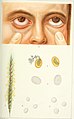
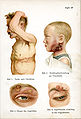
 Pediatrics. (1902) (14577803960).jpg 2,218 × 3,472; 856 KB
-
 PieIXretDiab.jpg 2,322 × 2,224; 923 KB
-
 Reke Salat.png 181 × 216; 50 KB
-
 Retrobulbarbleed.jpg 595 × 552; 102 KB
-
 Siebert 28.jpg 1,945 × 2,866; 1.13 MB
-
 Tape removal.jpg 818 × 615; 110 KB
-
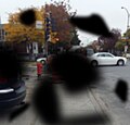
 Vision alterada por SOM diurno.jpg 1,289 × 780; 641 KB
-
 Vision alterada por SOM nocturno.jpg 1,063 × 672; 395 KB
-
 Yamai no Soshi - Eye Disease (part 1).jpeg 2,420 × 2,368; 3.19 MB
-
 Ülekoormuse põhjustatud paistetus.jpg 2,859 × 1,906; 3.3 MB
.jpg)


_(14596082218).jpg)

.png)
_Hrsg._von_W._Kolle_und_A._von_Wassermann_(1912-13)_(16662993341).jpg)


_(14577803960).jpg)


.jpeg)
