User talk:BruceBlaus
|
Our first steps tour and our frequently asked questions will help you a lot after registration. They explain how to customize the interface (for example the language), how to upload files and our basic licensing policy (Wikimedia Commons only accepts free content). You don't need technical skills in order to contribute here. Be bold when contributing and assume good faith when interacting with others. This is a wiki. More information is available at the community portal. You may ask questions at the help desk, village pump or on IRC channel #wikimedia-commons (webchat). You can also contact an administrator on their talk page. If you have a specific copyright question, ask at the copyright village pump. |
|
-- Wikimedia Commons Welcome (talk) 23:02, 23 July 2013 (UTC)
A barnstar for you![edit]

|
The Graphic Designer's Barnstar |
| Nice cell biology pictures! — Love, Kelvinsong talk 22:52, 6 September 2013 (UTC) |
File:Blausen 0330 EarAnatomy MiddleEar.png[edit]
Sorry I made a mistake with this file, when I just wanted to translate it into Spanish, I modified the original file wrongly. I have tried to unmake my edition but I'm not being able. Sorry again. Repair my mistake please, and tell me, if possible, how I can upload an image translated from Blausen. Thanks in advance. --Elboy99 (talk) 02:15, 1 December 2013 (UTC)
- Ok, I have been able at least to revert to the original file. Sorry for the inconvenience.--Elboy99 (talk) 02:15, 1 December 2013 (UTC)
Hello Elboy99. Thank you for reverting to the original file. Unfortunately, we cannot release layered files where the text can be changed; therefore there's no easy way to translate over the original English text. BruceBlaus (talk) 16:13, 3 December 2013 (UTC)BruceBlaus
Dear Bruce Blaus, I greatly admire your illustration, the pelvis: https://en.wikipedia.org/wiki/File:Blausen_0723_Pelvis.png . I wish to use it in our publication "Pediatric Surgery - Diagnosis and Treatment". I see that it has a Creative commons attribution 3.0 unported license, and will attribute it as such, but I wished to also notify you of my plans and request. The illustration will greatly enhance our chapter on pelvic fractures! Warm regards, Chris Coppola. Villac (talk) 00:56, 9 December 2013 (UTC)
Hello Villac. Yes, as long as we are attributed, the usage is fine. Thank you for letting me know. BruceBlaus (talk) 19:44, 11 December 2013 (UTC)BruceBlaus
Thank you very much, BruceBlausen! (for your art and your generosity!) I will attribute in this way: i. Figure: Pediatric Pelvis 1 – The pelvis consists of the pubis, the ischium, and the ileum. Adapted from Bruce Blaus under Creative Commons Attribution 3.0 Unported license as published on http://en.wikipedia.org/wiki/File:Blausen_0723_Pelvis.png . (I will edit this if you wish.) Merry Christmas! Villac (talk) 03:49, 14 December 2013 (UTC)
File:Blausen_0152_CABG_All.png[edit]
Dear Bruce Blaus, I was just reading https://en.wikipedia.org/wiki/Coronary_artery_bypass_surgery and noticed that in your - beautiful - picturehttps://en.wikipedia.org/wiki/File:Blausen_0152_CABG_All.png the triple and quadruple bypass graft have the exact same picture in use. As I see you allready have https://commons.wikimedia.org/wiki/File:Blausen_0154_CABG_Quadruple.png I figured it was a mistake and wanted to inform you on this. Kind regards Niekski (talk) 06:16, 30 April 2014 (UTC)
Thank you for letting us know Niekski. The image was fixed and re-uploaded this morning. BruceBlaus (talk) 13:36, 30 April 2014 (UTC)BruceBlaus
A barnstar for you![edit]

|
The Original Barnstar |
| Thank you for your excellent artwork and cooperative attitude! Villac (talk) 04:45, 18 December 2013 (UTC) |
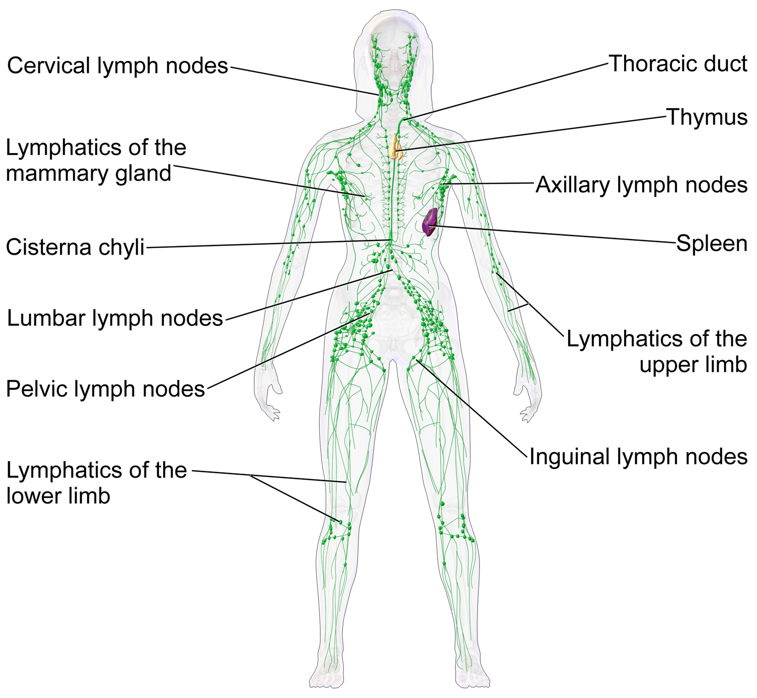
If you please, I have found another image I would love to use in our book: The Lymphatic System: https://upload.wikimedia.org/wikipedia/commons/f/f4/Blausen_0623_LymphaticSystem_Female.png I don't mean to pester you with requests, and answer only if you wish, but I will inform you when relying on your excellent work, always with attribution. Many thanks! Villac (talk) 04:47, 18 December 2013 (UTC) |} Hi Villac. Yes, as long as we are attributed for this image, the usage is fine. Thanks for letting me know. BruceBlaus (talk) 20:47, 3 January 2014 (UTC)BruceBlaus
permission to use images[edit]
Hello,
I would like to use 3 images that I found on Wikimedia Commons, created by Blausen Medical Communications.
These images will be used in offline e-learning training materials for healthcare workers in Tanzania. The materials will not be sold and no profit will be made from these materials - the usage is strictly educational. Please let me know if permission to use the materials is granted.
Images that we would like to use for this project are: http://commons.wikimedia.org/wiki/File:Blausen_0015_AllergicRhinitis.png, http://commons.wikimedia.org/wiki/File:Blausen_0390_EyeAnatomy_Sectional.png, and http://commons.wikimedia.org/wiki/File:Blausen_0389_EyeAnatomy_02.png
Many thanks.
Yes, as long as we are attributed as "Courtesy of Blausen.com", the 3 images may be used. BruceBlaus (talk) 21:51, 20 May 2014 (UTC)BruceBlaus
- Hey Bruce!
- I am about to publish a book about conception options and I would love to use your IUI image. The legal department at the publisher wanted me to double check with you that I could use it even though it will be in print. We will properly attribute the image to you and provide the website in print form in the endnotes. Is this OK for you?
- Thanks for creating this awesome image,
- Marea Mareagoodman (talk) 19:53, 24 August 2022 (UTC)
- Hello Marea,
- Thank you for contacting me. If the usage is non-profit, you may use the image free of charge in your publication with credit to Blausen in the endnotes. However, if the publication is for profit, the cost to use the illustration is $250.
- Thank you. You may contact us at: info@blausen.com to discuss. BruceBlaus (talk) 14:34, 25 August 2022 (UTC)
Thank you very much. We will attribute the images as instructed!
Publish gallery in Wikiversity Journal?[edit]
I see that you've gotten requests for permission to use images of yours in external works, and I believe there will be more requests in the future. Therefore, I think that your gallery is a excellent candidate for inclusion in Wikiversity Journal of Medicine. Thereby, other people using any image in external sources would be able to cite that image with a standard reference format, similarly to for example this image (as shown in its "Author" section). I can offer to prepare a gallery similar to Medical gallery of David Richfield 2014, using your uploads, and if you think it looks all right I can have it peer reviewed for inclusion in Wikiversity Journal. Does this sound interesting? If so, would it be ok to use the name "Blausen Medical Communications" as the creator? Mikael Häggström (talk) 18:56, 4 August 2014 (UTC)
Hello Mikael Häggström. We approve this request as long as we are cited as "Blausen.com" as the creator. Thank you! BruceBlaus (talk) 20:02, 4 August 2014 (UTC)BruceBlaus
- The images to be included in the journal are now on display at Wikiversity:Blausen gallery 2014, and the expected publication date is within a week. Images that are currently not included are given on the talk page at Wikiversity:Talk:Blausen gallery 2014 with explanations to such a decision. You may, if you wish, make an entry there to reply and/or motivate to have one or more of those images included in the publication as well. With a motivation, it is possible to have additional images included even after publication. Mikael Häggström (talk) 23:06, 24 August 2014 (UTC)
- The gallery is now published at Wikiversity:Blausen gallery 2014. Each of the included images can thereby now be cited as follows:
- Blausen.com. "Blausen gallery 2014". Wikiversity Journal of Medicine. DOI:10.15347/wjm/2014.010. ISSN 20018762.
- I will soon add this information in the image description pages. Mikael Häggström (talk) 18:03, 29 August 2014 (UTC)
- The gallery is now published at Wikiversity:Blausen gallery 2014. Each of the included images can thereby now be cited as follows:
Error in labelling in pic[edit]

Hi BruceBlaus,
I think there's an error in the labelling of File:Blausen 0861 Tonsils&Throat Anatomy2.png. I think the en:palatine tonsils have been mistakenly labelled as en:pharyngeal tonsils.
Cheers, Adrian J. Hunter (talk) 12:13, 9 September 2014 (UTC)
Hello Adrian J. Hunter. Thank you for the feedback, the image has been corrected. BruceBlaus (talk) 13:43, 9 September 2014 (UTC)BruceBlaus
- Great, thanks for the fix. Adrian J. Hunter (talk) 13:08, 19 September 2014 (UTC)
A barnstar for you![edit]
|
|
The Graphic Designer's Barnstar |
| Thanks for your wonderful illustrations! Jmwallach (talk) 15:46, 23 October 2014 (UTC) |
using images online[edit]
hello, I would like to know if images found under your user can be used on other websites, and in which conditions. Thanks!
Yes, as long as we are attributed as "Courtesy of Blausen.com" next to each image. Thank you, BruceBlaus (talk) 18:43, 14 January 2015 (UTC)BruceBlaus
Blausen_0406_FingerNailAnatomy.png *problem*[edit]
This illustration is misleading/wrong. The nail matrix is not just at the root. It extends under the nail all the way to the edge of the lunula. Thus, it is also misleading regarding the nail bed, which does not extend to the root? (File:Human_nail_anatomy.jpg is apparently better than File:Blausen_0406_FingerNailAnatomy.png) -71.174.183.177 20:53, 9 April 2015 (UTC)
We appreciate the feedback 71.174.183.177 . We revised the image to have a color gradient between the nail matrix (or germinal matrix) and the nail bed (or sterile matrix). The nail matrix is continuous with the nail bed. The leader lines are accurate when pointing to this anatomy. Thank you. BruceBlaus (talk) 22:06, 9 April 2015 (UTC)BruceBlaus
Use of an image of Blaus for a continuous medical education program[edit]
Dear Mr. Blaus,
I work for a pharmaceutical company and I am in the process of creating a Logo for our first continuous medical education event. This is a continuous medical education event co-sponsored by two pharmaceutical companies which might frequenty taking place in the future in Switzerland, depending on its success. I would like to use the following picture as part of the Logo. Of course I would need to remove the naming and adapt it a bit. I have already created a flyer for this event with a draft of the Logo. Is it possible to use your image for this event? In case you need more information please do not hesitate to contact me.
https://en.wikipedia.org/wiki/Neuron#/media/File:Blausen_0657_MultipolarNeuron.png
Best regards, Nadja
Hello Nadja (80.218.158.90), Unfortunately we cannot permit the usage of our content for logo purposes without paying a licensing fee. Please contact us at info@blausen.com for pricing information. Thank you 2606:A000:110A:C142:BCD1:FBFC:7245:F159 15:16, 24 July 2015 (UTC)bblaus
Request for permission to use image[edit]
Dear Bruce, I'm a publisher from Namibia who would like to use your image of Diffusion in one of my publications. I know that you have specified that the work is free, however, I need your explicit permission to be able to use this image.Please kindly contact me via email: malimap@nph.com.na to discuss this further. I look forward to hearing from you. Warm regards, Patrycja.
SVG (Scalable Vector Graphics)[edit]
| In other languages (translate this) |
|---|
|
|

|
Thank you for uploading some images!
Did you know that Wikimedia Commons recommends the SVG (Scalable Vector Graphics) format for certain types of images? Scalable Vector Graphics are designed to look appropriate at any scale, and SVG images are easier to modify and translate, helping Wikimedia to distribute knowledge to all of the world. A lot of modern programs support SVG export. If you encountered problems or have questions, don't hesitate to ask me, a member of the Graphic Lab, or the Graphics village pump. Uploading images in SVG format isn't mandatory, but it would help. (To avoid any misunderstandings, please don't just put raster images into an SVG container as embedded raster.) Thanks, and happy editing!
|
You have some awesome work! Keep it up - but SVG would be doubly awesome! :) Domdomegg (talk) 22:02, 22 January 2016 (UTC)
Image in vector or 300dpi format?[edit]
Thank you for this image: https://en.wikipedia.org/wiki/Facet_joint#/media/File:Facet_Joints.png. We like it so much we want to include it in our brochure, of course citing you. But we would love a print-ready, 300 dpi or vector version of this graphic to ensure it's quality in the brochure. Do you happen to have one we can purchase from you? Thanks so much!
- Hello there Kristindh. We appologise for taking so long to reply to your question. If you are still interested in the image, you can directly email us at info@blausen.com. BruceBlaus (talk) 20:46, 8 April 2016 (UTC)
SVG versions[edit]
I created an SVG version of this image for use in the article on tooth cavities. It has been deemed unsuitable. I tried. I am sure a photograph will be much better at depicting dentin tubules and enamel rods. I have requested that my SVG diagram be deleted. Check it out while you can 'cause its goin' fast! Or maybe you are gone from Commons. Wouldn't be the furst time. KDS4444 (talk) 09:53, 21 November 2016 (UTC)
Cystocele.png[edit]
Hello!. You should always try to categorize your uploaded media (ONLY) in a more accurate category. In this case Category:Cystocele. Similar problem in File:Dacryostenosis Blocked Tear Duct.png. Thanks. Jmarchn (talk) 06:58, 2 March 2017 (UTC)
Diabetes Foot Ulcers.png[edit]
Hello!. You should always try to categorize your uploaded media in a more accurate category. In this case Category:Diabetic foot ulcer. Thanks. Jmarchn (talk) 07:01, 2 March 2017 (UTC)
File:Intussusception.png[edit]
Hello!. You should always try to categorize your uploaded media in a more accurate category. In this case Category:Intussusception. Thanks. Jmarchn (talk) 15:47, 10 June 2017 (UTC)
File:Hemorrhoids.png[edit]
Hello!. You should always try to categorize your uploaded media in a more accurate category. In this case Category:Hemorrhoids. Thanks. Jmarchn (talk) 15:49, 10 June 2017 (UTC)
 |
File:Gamma Knife Surgery.png has been marked as a possible copyright violation. Wikimedia Commons only accepts free content—that is, images and other media files that can be used by anyone, for any purpose. Traditional copyright law does not grant these freedoms, and unless noted otherwise, everything you find on the web is copyrighted and not permitted here. For details on what is acceptable, please read Commons:Licensing. You may also find Commons:Copyright rules useful, or you can ask questions about Commons policies at the Commons:Help desk. If you are the copyright holder and the creator of the file, please read Commons:But it's my own work! for tips on how to provide evidence of that.
The file you added has been deleted. If you have written permission from the copyright holder, please have them send us a free license release via COM:VRT. If you believe that the deletion was not in accordance with policy, you may request undeletion. (It is not necessary to request undeletion if using VRT; the file will be automatically restored at the conclusion of the process.)
|
Wdwd (talk) 13:38, 18 February 2018 (UTC)
UR the GOAT![edit]
.jpg)
Greatest Of All Time!
LisaP12 (talk) 00:02, 27 July 2018 (UTC)
Neuron[edit]
Thanks for this. Tony (talk) 11:02, 2 February 2020 (UTC)
translate an image[edit]
Hi Blause, i'm an italian medical student and i want to ask you if i can use one of your image with the tranlation in italian for my final thesis. How can I show the modifications in the credits? thank you — Preceding unsigned comment added by Annanounce (talk • contribs) 18:58, 30 March 2021 (UTC)
Hi Blause, firstly, I would like to thank you for your work. Great indeed, educative and I can tell they are also entertaining, which is important. I like those vivid colours. Now there are few problems concerning some images at article on CABG. I am preparing it for a GAN, so I am wondering if you can do some improvements.
-
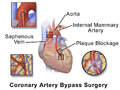 Illustration depicting coronary artery bypass surgery (double bypass)
 -
 Illustration of Single bypass
 -
 Illustration of Double bypass
 -
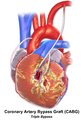 Illustration of Triple bypass
 -
 Illustration of Quadruple bypass


- File:Blausen 0151 CABG 02.png|Illustration depicting coronary artery bypass surgery (double bypass)
- error: LIMA is not sutured at left subclavian artery.
- LAD does not have any significant branches supplying Right Ventricle.
- Plaque blockage at LAT is red, should be white instead, which is the color of atheroma. As it is now, it looks as a thrombus or a clot.
- The saphnenous-RCA anastomosis is placed in a very awkward position- at the proximal RCA which is extremely rare.
- File:Blausen 0155 CABG Single.png|Illustration of Single bypass
- I coulndt understand what those two patches left and right of LAD represent. If they are necrotic areas, that is a wrong way to illustrate it and the anastomosis should be after the necrotic island, not before of it.
- Why LIMA changes its color as it gets close to LAD?
- Again, there should be no branches of LAD suppling right ventricle.
- File:Blausen 0153 CABG Double.png|Illustration of Double bypass
- The same question on those islands, and again the anastomosis of saphenous-RCA is too high.
- File:Blausen 0156 CABG Triple.png|Illustration of Triple bypass
- I liked that Y anastomosis but you forgot to place some stitches there! It will be leaking!
- File:Blausen 0154 CABG Quadruple.png|Illustration of Quadruple bypass
- The proximal anastomosis of the saphenous supplying Cx, is placed in the arch, whilst it should be sited at the front of the ascending aorta. Even the other proximal anastomosis (that supplies RCA) is a little bit too high.
Can you have a look at those details? Thank you in advance! Cinadon36 (talk) 08:36, 7 October 2022 (UTC)
PS-also, another comment. I think illustrating venous system of the heart, being a little confusing and might be factually wrong, as it follows the logic that for a given artery, there is a vein, which is not exactly the case in the heart. I think it would be better to remove them.Cinadon36 (talk) 08:42, 7 October 2022 (UTC)
- Hello Cinadon, we have updated the illustrations to more accurately represent anastomoses and necrosis, as well as include labels, as well as updating the overall look! We hope these meet your approval. BruceBlaus (talk) 23:49, 7 December 2022 (UTC)
I'll attribute your Wikipedia account for your excellent tritium model.[edit]
I'm borrowing the image for a project, I'll attribute your Wikipedia account as a watermark.
RealNamesAreFineZ.E.O (talk) 11:30, 1 September 2023 (UTC)
|
File:Discogenic Pain.png has been marked as a possible copyright violation. Wikimedia Commons only accepts free content—that is, images and other media files that can be used by anyone, for any purpose. Traditional copyright law does not grant these freedoms, and unless noted otherwise, everything you find on the web is copyrighted and not permitted here. For details on what is acceptable, please read Commons:Licensing. You may also find Commons:Copyright rules useful, or you can ask questions about Commons policies at the Commons:Help desk. If you are the copyright holder and the creator of the file, please read Commons:But it's my own work! for tips on how to provide evidence of that.
The file you added may soon be deleted. If you have written permission from the copyright holder, please replace the copyvio tag with {{subst:OP}} and have them send us a free license release via COM:VRT. If you disagree that the file is a copyright violation for any other reason, please replace the copyvio tag with a regular deletion request.
|
User who nominated the file for deletion (Nominator) : Gnomingstuff.
I'm a computer program; please don't ask me questions but ask the user who nominated your file(s) for deletion or at our Help Desk. //Deletion Notification Bot 2 (talk) 06:48, 24 December 2023 (UTC)