File:Perivascular-Mural-Cells-of-the-Mouse-Choroid-Demonstrate-Morphological-Diversity-That-Is-pone.0053386.s004.ogv
Jump to navigation
Jump to search
Size of this JPG preview of this OGG file: 305 × 600 pixels. Other resolutions: 122 × 240 pixels | 244 × 480 pixels | 390 × 767 pixels | 521 × 1,024 pixels | 2,048 × 4,026 pixels.
Original file (Ogg Theora video file, length 5.5 s, 2,048 × 4,026 pixels, 3.35 Mbps, file size: 2.2 MB)
Captions
Captions
Add a one-line explanation of what this file represents
Summary[edit]
| Description |
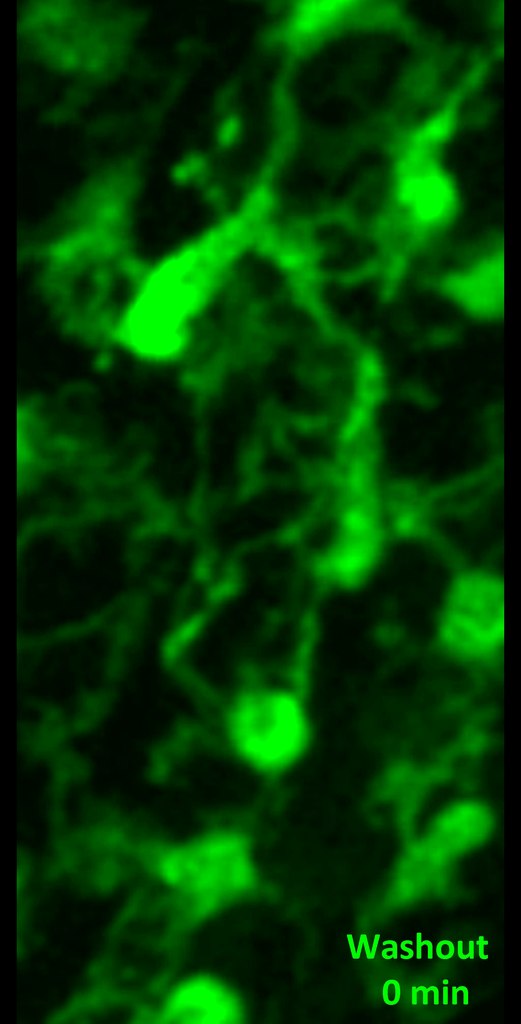
English: Response of a choroidal arteriole (type 3 vessel) before and at different time points after the application of calcium ionophore A23187 (10 µM) in the recording medium. Constriction of the vessel was observed 5 minutes after A23187application and sustained for the 30 minutes of A23187 application. Vascular constriction was long-lasting and only fractionally reversed 60 minutes after washout. |
||
| Date | |||
| Source | Movie S3 from Condren A, Kumar A, Mettu P, Liang K, Zhao L, Tsai J, Fariss R, Wong W (2013). "Perivascular Mural Cells of the Mouse Choroid Demonstrate Morphological Diversity That Is Correlated to Vasoregulatory Function". PLOS ONE. DOI:10.1371/journal.pone.0053386. PMID 23308209. PMC: 3537675. | ||
| Author | Condren A, Kumar A, Mettu P, Liang K, Zhao L, Tsai J, Fariss R, Wong W | ||
| Permission (Reusing this file) |
|
||
| Provenance |
|
File history
Click on a date/time to view the file as it appeared at that time.
| Date/Time | Thumbnail | Dimensions | User | Comment | |
|---|---|---|---|---|---|
| current | 02:03, 3 June 2013 | 5.5 s, 2,048 × 4,026 (2.2 MB) | Open Access Media Importer Bot (talk | contribs) | Automatically uploaded media file from Open Access source. Please report problems or suggestions here. |
You cannot overwrite this file.
File usage on Commons
There are no pages that use this file.
Transcode status
Update transcode statusMetadata
Categories:
- Vascular system of the eyes
- Mus musculus eyes
- Microscopic images of Mus musculus circulatory system
- Cell physiology
- Immunologic techniques
- Immunohistochemical analysis
- Animal models of disease
- Molecular cell biology
- Videos of cell types
- Muscle cells
- Videos of cardiovascular system
- Macular disorders
- Videos of disorders of choroid and retina
- Media from PLOS ONE