File:PBP catalysis.svg

Original file (SVG file, nominally 1,142 × 1,567 pixels, file size: 1.44 MB)
Captions
Captions
Summary[edit]
| Description |
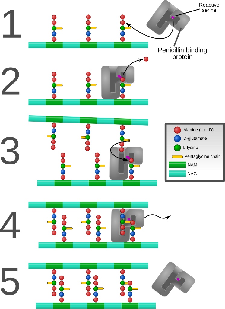
English: Diagram depicting formation of cross-links in the bacterial cell wall by a penicillin binding protein (PBP, an enzyme).
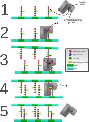
1. The bacterial cell wall consists of strands of repeating N-acetylglucosamine (NAG) and N-acetylmuramic acid (NAM) subunits. The NAM subunits have short peptide chains attached to them. (The exact composition of these can vary. The proximal alanine is usually L-ala and the distal two are usually D-ala.) These chains, in turn, are bound to chains of 5 glycine residues that will be used in cross-linking. 2. The PBP forms a bond with the peptide side chain at the second most distal alanine residue. This displaces the most distal alanine residue. 3. Another strand of bacterial cell wall arrives. The free end of one of the pentaglycine chains displaces the PBP and forms a bond with the terminal alanine on the other strand. 4. After being displaced, the PBP diffuses away. 5. The formation of one cross-link is complete. |
| Date | |
| Source | Own work |
| Author | Mcstrother |
| Other versions |
[]
|


Licensing[edit]
- You are free:
- to share – to copy, distribute and transmit the work
- to remix – to adapt the work
- Under the following conditions:
- attribution – You must give appropriate credit, provide a link to the license, and indicate if changes were made. You may do so in any reasonable manner, but not in any way that suggests the licensor endorses you or your use.
File history
Click on a date/time to view the file as it appeared at that time.
| Date/Time | Thumbnail | Dimensions | User | Comment | |
|---|---|---|---|---|---|
| current | 19:26, 9 September 2011 | | 1,142 × 1,567 (1.44 MB) | Mcstrother (talk | contribs) | Major revision. Corrected inaccuracies in previous image. |
| 04:15, 3 May 2011 |  | 1,139 × 1,062 (850 KB) | Mcstrother (talk | contribs) | Changed all fonts to Liberation Sans | |
| 03:46, 10 April 2011 |  | 1,139 × 1,062 (850 KB) | Mcstrother (talk | contribs) | Changed color of carbohydrate chain. | |
| 03:30, 7 March 2011 |  | 1,139 × 1,062 (835 KB) | Mcstrother (talk | contribs) | {{Information |Description ={{en|1=Diagram depicting formation of cross-links in the bacterial cell wall by a penicillin binding protein (PBP, an enzyme). 1. The bacterial cell wall consists of strands of repeating N-acetylglucosamine (NAG) and N-ace |
You cannot overwrite this file.
File usage on Commons
The following 5 pages use this file:
File usage on other wikis
The following other wikis use this file:
- Usage on en.wikipedia.org
- Usage on es.wikipedia.org
- Usage on fa.wikipedia.org
- Usage on ga.wikipedia.org
- Usage on gl.wikipedia.org
- Usage on hu.wikipedia.org
- Usage on it.wikipedia.org
- Usage on mk.wikipedia.org
- Usage on th.wikipedia.org