Category:Enzyme reactions
Jump to navigation
Jump to search
Subcategories
This category has the following 6 subcategories, out of 6 total.
H
- Hydrolases reactions (20 F)
I
- Isomerases reactions (15 F)
L
- Lyases reactions (15 F)
M
O
T
- Transferases reactions (16 F)
Media in category "Enzyme reactions"
The following 200 files are in this category, out of 542 total.
(previous page) (next page)-
 062821 Chymotrypsin Tetrahedral Intermediates.pdf 875 × 443; 845 KB
-
 1 6glucosidase reaction.svg 681 × 245; 23 KB
-
 17a-hydroxyprogesterone reaction with 21-hydroxylase.png 586 × 244; 32 KB
-
 17a-progesterone reaction with 21-hydroxylase.png 593 × 241; 31 KB
-
 2368fig1.jpg 720 × 111; 12 KB
-
 2OG Dioxygenase-Catalysed Reaction.png 1,075 × 145; 10 KB
-
 3-ketoactl-ACP synthetase.png 594 × 221; 34 KB
-
 3MBCG to MMB degradation.jpg 982 × 147; 16 KB
-
 4-(2-carboxyphenyl)-4-oxobutanoyl-CoA to 1,4-dihydroxy-2-naphthoyl-CoA.png 1,840 × 552; 16 KB
-
 5-HTP decarboxylation labeled.svg 534 × 108; 17 KB
-
 ABA-GE1.JPG 505 × 532; 24 KB
-
 Abysommicin c PABA pathway.png 614 × 343; 57 KB
-
 ACAC mechanism.png 627 × 234; 7 KB
-
 Acetoacetate to beta-hydroxybutyrate.svg 1,052 × 400; 23 KB
-
 Acetolactat-Synthase-reaction.svg 360 × 176; 4 KB
-
 Acety-CoA ACP transacylase.png 546 × 113; 15 KB
-
 Acetylation, reaction.gif 1,631 × 930; 47 KB
-
 Aconitase Tritium Crossover Scheme.png 3,057 × 584; 116 KB
-
 Acrosin Catalytic Mechanism.jpg 2,012 × 912; 123 KB
-
 ACS paramagetic and diamagnetic mechanisms.jpg 425 × 342; 22 KB
-
 Activation of fatty acids (zh-cn).svg 1,500 × 400; 41 KB
-
 Activation of fatty acids jp.svg 1,993 × 394; 219 KB
-
 Active Site 1 - 2.png 574 × 291; 8 KB
-
 Active Site 2.png 574 × 291; 9 KB
-
 Active site mechanism of mercuric reductase.png 617 × 351; 12 KB
-
 Acylphosphate rxn.png 3,211 × 824; 32 KB
-
 Acylphosphate rxn.svg 484 × 124; 19 KB
-
 ADA 2.jpg 674 × 228; 35 KB
-
 ADAR reaction.jpg 354 × 123; 11 KB
-
 Adenine deaminase scheme.jpg 200 × 290; 8 KB
-
 ADH formule.png 2,198 × 1,618; 163 KB
-
 Aequorin mechanism.png 3,063 × 2,672; 63 KB
-
 Aging of Soman in ChemDraw.png 339 × 100; 3 KB
-
 ALA-synthesis-from-glutamyl-tRNA.png 1,656 × 402; 9 KB
-
 ALA-synthesis-from-succinyl-CoA.png 1,130 × 316; 6 KB
-
 Alanin aminotranszferáz.png 647 × 213; 10 KB
-
 Alanine aminotransférase (ALAT).png 2,132 × 1,632; 214 KB
-
 Alanine transaminase reaction.PNG 2,384 × 690; 54 KB
-
 Aldehyde dehydrogenase mechanism.png 2,114 × 432; 28 KB
-
 Aldehyde dehydrogenase mechanism.svg 780 × 129; 93 KB
-
 Allelopathie bei Juglans regia.png 690 × 542; 20 KB
-
 Alpha oxidation part I.svg 523 × 98; 31 KB
-
 Alpha oxidation part II.svg 670 × 234; 39 KB
-
 Alpha oxidation part III.svg 579 × 118; 25 KB
-
 Alpha oxidation part IV.svg 460 × 119; 23 KB
-
 Alpha oxidation part V.svg 460 × 124; 31 KB
-
 Amine reaction.png 440 × 160; 3 KB
-
 Aminoacyladenylat.svg 1,494 × 346; 81 KB
-
 Aminoacylase.png 1,161 × 237; 14 KB
-
 Aminolevulinic synthesis.png 2,517 × 851; 26 KB
-
 Amylase hydrolysisl 1-4.png 432 × 436; 31 KB
-
 Amylase reaction.png 575 × 428; 46 KB
-

-
 AnPRT catalyzed rxn 1.png 1,084 × 564; 47 KB
-
 AOX reaction.png 323 × 70; 4 KB
-
 APRTase Reaction Scheme.png 467 × 125; 13 KB
-
 ARPTase Reaction Scheme.png 467 × 120; 13 KB
-
 Arylsulfatase B reaction in dermatan sulfate degradation.png 350 × 590; 30 KB
-
 Arylsulfatase B reaction.png 1,162 × 342; 32 KB
-
 ASPA mechanism.jpg 956 × 221; 33 KB
-
 ASPA overall reaction 2.jpg 704 × 148; 18 KB
-
 ASPA overall reaction.jpg 604 × 163; 16 KB
-
 Asparagine synthase rn.png 1,128 × 226; 8 KB
-
 Aspartate aminotransferase reaction.gif 624 × 128; 3 KB
-
 Aspartate aminotransferase reaction.png 718 × 90; 3 KB
-
 Aspartate transaminase reaction.svg 620 × 276; 53 KB
-
 Aspartate transaminase rn.png 1,405 × 297; 10 KB
-
 Aspartyl proteases.png 1,355 × 1,328; 1.2 MB
-
 Assim Sulfate Reduction.png 1,266 × 575; 109 KB
-
 ATCase reaction.jpg 515 × 230; 41 KB
-
 ATCase Reaction.jpg 1,319 × 691; 85 KB
-
 B lyase.gif 1,053 × 244; 8 KB
-
 B to o.png 4,491 × 2,293; 380 KB
-
 B-Glu4.png 584 × 162; 7 KB
-
 B-Raf Phosphorylation Mechanism.png 1,056 × 995; 72 KB
-
 BCIP reaction.png 4,412 × 1,015; 37 KB
-
 BCIP reaction.svg 727 × 140; 51 KB
-
 BCKDC catalytic mechanism step 1.png 942 × 522; 62 KB
-
 Before and after ELF97.png 263 × 413; 21 KB
-
 Beta cystathionine synthase.svg 838 × 255; 105 KB
-
 Beta oxidation of unsaturated fatty acid (zh-cn).svg 490 × 800; 95 KB
-
 Beta oxidation of unsaturated fatty acid jp.svg 490 × 800; 292 KB
-
 Beta-ketothiolase.png 814 × 880; 62 KB
-
 Beta-lactam closure routes.png 2,176 × 1,368; 53 KB
-
 Beta-LAS.jpg 513 × 576; 66 KB
-
 Beta-Oxidation 1 jp.svg 771 × 149; 70 KB
-
 Beta-Oxidation 2 jp.png 733 × 124; 21 KB
-
 Beta-Oxidation 2 jp.svg 771 × 149; 80 KB
-
 Beta-Oxidation 3 jp.png 696 × 135; 23 KB
-
 Beta-Oxidation 3 jp.svg 732 × 160; 96 KB
-
 Beta-Oxidation 4 jp.png 745 × 102; 20 KB
-
 Beta-Oxidation 4 jp.svg 800 × 120; 104 KB
-
 Beta-Oxidation1.svg 771 × 149; 57 KB
-
 Beta-Oxidation2.svg 772 × 143; 64 KB
-
 Beta-Oxidation3.svg 771 × 169; 73 KB
-
 Beta-Oxidation4.svg 971 × 134; 59 KB
-
 Bilirubin dönüşüm şeması.png 1,208 × 1,400; 93 KB
-
 Biochemistry metabolism 5c.png 647 × 141; 13 KB
-
 Bioluminescence reaction.jpg 984 × 113; 30 KB
-
 Biosynthese von N-Carbamoylaspartat.svg 915 × 215; 189 KB
-
 Biosynthesis of luciferin Luciola lateralis.svg 512 × 263; 53 KB
-
 Butyrate Kinase Mechanism.jpg 437 × 290; 22 KB
-
 BVR mechanism pl.png 3,938 × 1,484; 41 KB
-
 BVR mechanism.png 3,938 × 1,484; 80 KB
-
 C-di-AMP synthesis and degradation reaction.png 2,934 × 1,234; 374 KB
-
 Calcein-AM.svg 512 × 731; 46 KB
-
 Captura de pantalla 2017-10-25 a les 22.00.01.png 921 × 470; 95 KB
-
 Carboxypeptidase catalysis (zh-cn).svg 350 × 200; 43 KB
-
 Carboxypeptidase catalysis (zh-tw).svg 550 × 300; 50 KB
-
 Carboxypeptidase-anhydride mechanism.png 548 × 416; 32 KB
-
 Carboxypeptidase.jpg 614 × 481; 33 KB
-
 Carnitine Acetyltransferase Mechanism.tiff 762 × 354; 48 KB
-
 Carnitine carrier system.svg 620 × 775; 172 KB
-
 Carnitinshuttle.png 3,348 × 2,974; 52 KB
-
 Catalase reaction.png 168 × 142; 2 KB
-
 Catalytic mechanisms of GNAT and MYST family HATs..png 1,317 × 845; 146 KB
-
 Catechol dioxygenase reaction.svg 1,175 × 213; 17 KB
-
 Catecol-Quinona.svg 405 × 102; 15 KB
-
 CEA synthase mechanism.png 1,960 × 1,816; 52 KB
-
 CellularTrxR.png 1,241 × 285; 19 KB
-
 Chalcone isomerase.svg 1,515 × 385; 27 KB
-
 Chemdraw wikipedia.tif 2,442 × 622; 84 KB
-
 Chemdrawfinalfinalmary.jpg 2,764 × 238; 48 KB
-
 Chemical glycosylation FC fragment.png 1,464 × 874; 374 KB
-
 Chitosan Synthese.svg 712 × 594; 110 KB
-
 Chlorinase reaction.png 1,629 × 255; 27 KB
-
 Cholesterol-Synthesis-Reaction10.png 3,453 × 354; 19 KB
-
 Chorismate Mutase Scheme.png 1,334 × 489; 8 KB
-
 Chorismate pathway 2.png 1,320 × 329; 11 KB
-
 Citrulline metabolism.png 2,675 × 705; 166 KB
-
 Cleave side chain.png 2,545 × 483; 29 KB
-
 Cocarboxylase.svg 512 × 174; 71 KB
-
 Conversao.png 459 × 339; 61 KB
-
 Conversion by COQ5.svg 604 × 241; 39 KB
-
 Cortisone Reductase Deficiency effects on HPA and body.png 1,198 × 724; 123 KB
-
 Coupled assay tr.png 594 × 216; 10 KB
-
 Coupling of two propionyl-units.jpg 675 × 313; 25 KB
-
 Creatine kinase rxn.png 2,408 × 620; 43 KB
-
 Cyanase enzymatic reaction scheme.png 1,018 × 270; 39 KB
-
 Cystathionine gamma-synthase.svg 512 × 74; 19 KB
-
 Cysteinprotease Peptidspaltung.svg 1,368 × 1,548; 105 KB
-
 Cysteinproteasen ; Spaltung einer Peptidbindung durch eine Cysteinprotease.svg 1,368 × 1,548; 102 KB
-
 D-LDH reaction.svg 620 × 211; 21 KB
-
 DAHPtoG3P.png 682 × 183; 4 KB
-
 DC général.png 1,077 × 341; 28 KB
-
 De-Novo-ODCase.PNG 456 × 171; 5 KB
-
 De-Novo-OPRT-ase.PNG 675 × 174; 7 KB
-
 Decomposition.png 2,348 × 645; 87 KB
-
 Decomposição pela Linamarase.png 938 × 273; 13 KB
-
 Degradation of Isethionate Catalyzed by IseG.jpg 980 × 127; 33 KB
-
 Degradation of uric acid.svg 820 × 403; 67 KB
-
 Demethylation of Lanosterol.png 1,058 × 914; 65 KB
-
 Demethylering van SAM-page-001.jpg 1,240 × 1,175; 80 KB
-
 DHFR Reaction Scheme.png 721 × 427; 23 KB
-
 DHFR rxn.svg 736 × 757; 44 KB
-
 Dihydropteroat-Synthase - Reaktion.svg 1,895 × 461; 81 KB
-
 Dihydroxyacetone phosphate to glycerol 3-phosphate en.svg 2,980 × 1,300; 27 KB
-
 Dihydroxyacetone phosphate to glycerol 3-phosphate es.svg 2,980 × 1,300; 27 KB
-
 Dihydroxyacetone phosphate to glycerol 3-phosphate zh.svg 2,980 × 1,300; 27 KB
-
 DNase MOA.jpg 866 × 686; 52 KB
-
 Dopamine beta-hydroxylase reaction.png 726 × 412; 35 KB
-
 Dopamine beta-hydroxylase reaction.svg 583 × 196; 14 KB
-
 Dopamine beta-hydroxylase.png 663 × 331; 33 KB
-
 Dopamine beta-monooxygenase reaction.svg 583 × 196; 14 KB
-
 DPAGT1 Catalysis.tif 1,280 × 720; 122 KB
-
 EC 2.1.1.115 rxn.png 3,800 × 1,514; 293 KB
-
 EC 5.3.1.16 reaction.PNG 2,114 × 723; 46 KB
-
 Endochitinase.png 1,900 × 571; 142 KB
-
 Energia12.png 640 × 360; 3 KB
-
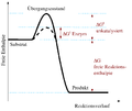 Energiediagramm Enzymreaktion.png 976 × 823; 68 KB
-
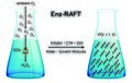 Enz-RAFT schematic.jpg 2,096 × 1,333; 1.25 MB
-
 Enzymatic oxidation of arachidonic acid.png 525 × 195; 24 KB
-
 EnzymatischeAktivitätHNL.svg 677 × 145; 36 KB
-
 Enzyme activation energy.png 314 × 294; 16 KB
-
 Enzyme mechanism 1.jpg 657 × 254; 32 KB
-
 Enzyme realsse of preprobe.jpg 3,000 × 2,250; 226 KB
-
 Enzyme transition state.jpg 430 × 193; 8 KB
-
 EPSPreactionII.svg 1,483 × 325; 79 KB
-
 Erwinaze reaction.png 415 × 202; 10 KB
-
 Esquema 4S Pathway.svg 512 × 360; 958 KB
-
 EsteraseScope3.png 370 × 144; 16 KB
-
 EsteraseScope4.png 576 × 137; 15 KB
-
 EsteraseScope5.png 337 × 91; 12 KB
-
 EsteraseScope6.png 691 × 85; 14 KB
-
 EsteraseScope7.png 587 × 140; 15 KB
-
 EsteraseSynth1.png 578 × 229; 24 KB
-
 EsteraseSynth2.png 743 × 258; 29 KB
-
 EsteraseSynth3.png 797 × 122; 20 KB
-
 Estriol synthesis.svg 1,640 × 365; 36 KB
-
 Exogenous mechanism of Argpyrimidine.png 509 × 126; 14 KB
-
 Exophosphatase mechanism.png 1,708 × 1,041; 93 KB
-
 FAO.png 250 × 310; 27 KB
-
 FattyAcid-MB-OxidationByNAD.png 658 × 111; 3 KB
-
 Fementação alcoolica.jpg 335 × 551; 34 KB
-
 FGE mechansim.tif 1,272 × 2,466; 98 KB
-
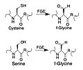 FGE serine-cysteine.jpg 646 × 546; 78 KB
-
 Figura 1.jpg 337 × 250; 9 KB
-
 Figura2.jpg 586 × 684; 33 KB
-
 FINAL GLUTAMATE.GIF 997 × 361; 8 KB
-
 Flavin Hydroperoxide Corrected.jpg 892 × 435; 34 KB


.svg)


.svg)
.svg)


-4-oxobutanoyl-CoA_to_1,4-dihydroxy-2-naphthoyl-CoA.png)
.svg)
.png)