File:Ion-Abrasion-Scanning-Electron-Microscopy-Reveals-Surface-Connected-Tubular-Conduits-in-HIV-ppat.1000591.s001.ogv
Jump to navigation
Jump to search
Size of this JPG preview of this OGG file: 605 × 599 pixels. Other resolutions: 242 × 240 pixels | 622 × 616 pixels.
Original file (Ogg multiplexed audio/video file, Theora/Vorbis, length 32 s, 622 × 616 pixels, 1.52 Mbps overall, file size: 5.72 MB)
Captions
Captions
Add a one-line explanation of what this file represents
Summary[edit]
| Description |
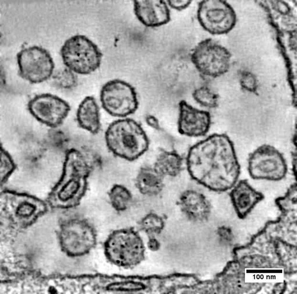
English: Slices through a dual-axis tomogram obtained from 150 nm thick cell sections cur from a fixed, stained, plastic-embedded block containing HIV-1 infected macrophages. Primary human monocyte-derived macrophages (MDM) were infected for 7 days with the primary isolate HIV-1 BaL. The region of the cell that can be explored is limited by the thickness of the section. |
||
| Date | |||
| Source | Video S1 from Bennett A, Narayan K, Shi D, Hartnell L, Gousset K, He H, Lowekamp B, Yoo T, Bliss D, Freed E, Subramaniam S (2009). "Ion-Abrasion Scanning Electron Microscopy Reveals Surface-Connected Tubular Conduits in HIV-Infected Macrophages". PLOS Pathogens. DOI:10.1371/journal.ppat.1000591. PMID 19779568. PMC: 2743285. | ||
| Author | Bennett A, Narayan K, Shi D, Hartnell L, Gousset K, He H, Lowekamp B, Yoo T, Bliss D, Freed E, Subramaniam S | ||
| Permission (Reusing this file) |
|
||
| Provenance |
|
File history
Click on a date/time to view the file as it appeared at that time.
| Date/Time | Thumbnail | Dimensions | User | Comment | |
|---|---|---|---|---|---|
| current | 13:01, 17 November 2012 | 32 s, 622 × 616 (5.72 MB) | Open Access Media Importer Bot (talk | contribs) | Automatically uploaded media file from Open Access source. Please report problems or suggestions here. |
You cannot overwrite this file.
File usage on Commons
There are no pages that use this file.