Category:Yolk sacs
Jump to navigation
Jump to search
membranous sac attached to an embryo, formed by cells of the hypoblast adjacent to the embryonic disk  | |||||
| Upload media | |||||
| Instance of |
| ||||
|---|---|---|---|---|---|
| Subclass of |
| ||||
| Made from material | |||||
| |||||
Media in category "Yolk sacs"
The following 115 files are in this category, out of 115 total.
-
 A close-up view of the same zebrafish.jpg 2,048 × 1,536; 1.23 MB
-
 A human embryo of 2 mm. in median sagittal section.jpg 838 × 1,006; 310 KB
-
 A zebrafish with congenital malformation of the yolk sac.jpg 2,048 × 1,536; 901 KB
-

-
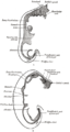
 Alimentary Canal during the 5th week.jpg 819 × 646; 218 KB
-
 Alimentary Canal in a human embryo of the 3rd week.jpg 967 × 624; 250 KB
-
 Allantois bird (01).jpg 1,350 × 1,093; 407 KB
-
 Allantois bird.jpg 1,062 × 1,674; 366 KB
-
 Atlantic salmon redd.jpg 1,145 × 749; 391 KB
-

-
-
 Cephalopods three with a very small yolk-sack.jpg 1,207 × 737; 697 KB
-
 Chicken embryo of about five days incubation.jpg 1,218 × 750; 1,003 KB
-
 Chicken embryo of about fourteen days incubation.jpg 1,216 × 736; 901 KB
-
-
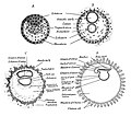
 Diagrams and images of human embryos at the gastrula stage.png 3,128 × 3,193; 804 KB
-
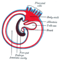
 Diagrams showing the development of the amnion, chorion and allantois.jpg 1,269 × 1,151; 605 KB
-
 Differentiation of the mesoderm in holoblastic and meroblastic types of development.jpg 1,617 × 1,467; 1,022 KB
-
 Diversity of vertebrate gastrulation.jpg 1,073 × 1,021; 415 KB
-
 Egg -chick -yolk sac-6a.jpg 613 × 1,024; 85 KB
-
 Embryo at 4 to 5 weeks fallopian tube (01).jpg 2,000 × 1,334; 1.35 MB
-
 Embryology (1949) (21285693065).jpg 1,035 × 1,115; 581 KB
-

-
 Foetus cat (01).jpg 954 × 612; 497 KB
-
 Foetus cat.jpg 667 × 827; 532 KB
-
 Formation of the Umbilical Region. human embryo, 1.7 mm. long.jpg 1,065 × 857; 974 KB
-
 Formation of the Umbilicus and Allantois. human embryo, 0.7 mm. long..jpg 1,152 × 803; 970 KB
-
 Formation- of the Umbilicus in an Embryo 2.5 mm.jpg 788 × 837; 598 KB
-
 Four diagrams showing hypothetical stages of early human embryos.jpg 1,631 × 1,434; 943 KB
-
 Frog embryo Sagittal section (2).jpg 782 × 694; 424 KB
-
 Frog embryo Sagittal section.jpg 850 × 745; 483 KB
-
 Frog's embryo transverse section.jpg 844 × 735; 549 KB
-
 Gastrulation forms in vertebrates.jpeg 1,280 × 1,173; 88 KB
-
 GR&PGCs.png 1,377 × 688; 187 KB
-
 Gray22.png 300 × 303; 23 KB
-
 Gray24.svg 252 × 161; 8 KB
-
 Gray25.svg 300 × 295; 13 KB
-
 Gray26.svg 344 × 345; 21 KB
-
 Gray27.png 300 × 298; 10 KB
-
 Gray28.svg 396 × 455; 14 KB
-
 Gray30.png 500 × 537; 74 KB
-
 Gray982.png 374 × 700; 25 KB
-
 Hatched trout eggs (6601049329).jpg 3,072 × 2,304; 1.86 MB
-
 Human embryo Section of embryonic rudiment in Peters' ovum (first week).jpg 1,141 × 857; 540 KB
-
 Human Yolk Sac from Tubal Pregnancy (32847892157).jpg 1,946 × 1,887; 891 KB
-
 Human Yolk Sac from Tubal Pregnancy (33914344418).jpg 1,670 × 2,559; 676 KB
-
 Human Yolk Sac from Tubal Pregnancy (33914344548).jpg 1,771 × 2,516; 1.06 MB
-
 Human Yolk Sac from Tubal Pregnancy (33914345538).jpg 1,643 × 2,167; 669 KB
-
 Human Yolk Sac from Tubal Pregnancy (47002175724).jpg 2,096 × 2,274; 1.02 MB
-
 Human Yolk Sac from Tubal Pregnancy (47002176474).jpg 2,026 × 1,962; 1.16 MB
-
 Human Yolk Sac from Tubal Pregnancy (47739058042).jpg 1,744 × 2,647; 1.35 MB
-
 Human Yolk Sac from Tubal Pregnancy (47739058172).jpg 2,330 × 2,384; 1.55 MB
-
 Human Yolk Sac from Tubal Pregnancy (47791378311).jpg 1,514 × 2,075; 527 KB
-
 Human Yolk Sac in Tubal Pregnancy (14961233588).jpg 2,048 × 1,536; 710 KB
-
 Human- Embryo, about 3.5 mm. long.jpg 803 × 791; 701 KB
-
 Human- Embryo, about 5 mm. long.jpg 825 × 846; 809 KB
-
 Ichthyologie; ou, Histoire naturelle des poissons (Plate 75) (6918354674).jpg 2,283 × 1,342; 392 KB
-
 Koorumata vikerforell.tif 3,840 × 2,880; 31.78 MB
-
 Koorunud vikerforell (Oncorhynchus mykiss).tif 3,840 × 2,880; 31.79 MB
-
 Latimeria chalumnae embryo.jpg 1,200 × 720; 286 KB
-
 Loligo advanced embryo.jpg 515 × 862; 500 KB
-
 Loligo ovum mesenteric cavity.jpg 912 × 578; 759 KB
-

-

-
-
 Mouse embryo Cellular expansion and morphology of CSF1R+ progenitors.jpg 1,983 × 1,946; 2.73 MB
-
 Mouse embryo Intravascular trafficking is independent of MYB and CX3CR1.jpg 1,360 × 2,512; 1.84 MB
-
 Mouse embryo Intravascular trafficking of CX3CR1+ YS pre-macrophages.jpg 1,780 × 2,460; 2.62 MB
-
 Mouse embryo Pre-macrophages infiltrate embryonic tissues.jpg 1,346 × 2,383; 2.14 MB
-
 Mouse embryo Trafficking is associated with cellular morphology.jpg 1,578 × 1,923; 1.16 MB
-
 Mouse embryo Trafficking kinetics of CSF1R+ cells are similar to pre-macrophages.jpg 1,790 × 1,508; 1.34 MB
-
 Mouse embryo Trafficking of KIT+ EMPs.jpg 1,358 × 1,888; 1.2 MB
-
 Mustelus antarcticus juvenile.jpg 1,200 × 639; 202 KB
-
 Newborns (12906215023).jpg 960 × 720; 143 KB
-
 Northern Pike Egg (7441919980).jpg 1,625 × 1,486; 367 KB
-
 Notopterus notopterus (10.3897-zse.93.13341) Figure 9.jpg 1,949 × 1,931; 1.95 MB
-

-
 Oncorhynchus gorbuscha larva.jpg 4,032 × 3,024; 2.88 MB
-
 Photo of the Week - Atlantic Salmon Sac Fry (5197995371).jpg 1,193 × 814; 728 KB
-
 Photo of the Week - Newly hatched bog turtle (4924259588).jpg 4,752 × 3,168; 7.07 MB
-
 Pseudorasbora parva (10.3897-zoologia.35.e22162) Figures 2–39.jpg 1,997 × 1,494; 1.42 MB
-
 Salmon newborn.jpg 800 × 600; 92 KB
-
 Salmonlarvakils 2.jpg 1,820 × 1,820; 449 KB
-
 Salmonlarvakils.jpg 1,249 × 1,821; 178 KB
-
 Sand devil embryo.jpg 150 × 117; 16 KB
-
 Sepia three late stages in the development.jpg 867 × 770; 309 KB
-

-
 The same zebrafish at a different angle.jpg 2,048 × 1,536; 1.22 MB
-
 Tubal Pregnancy with Yolk Sac Inside Chorionic Cavity (14961233838).jpg 2,048 × 1,536; 720 KB
-
 Umbilical Cord of a Human Embryo 12.5 mm. in length.jpg 1,001 × 1,445; 1.15 MB
-
 Umbilical Region in a Human Embryo 23 mm. in length.jpg 960 × 833; 729 KB
-
 Umbilical Region of a Human Embryo 10 mm. in length.jpg 1,167 × 957; 1.09 MB
-
 Umbilical Region of a Human Embryo 3 cm. long.jpg 804 × 769; 769 KB
-
 Umbilical Region, the Cord, and the Placenta at Term.jpg 778 × 635; 577 KB
-
 Yolk sac human.jpg 1,054 × 1,355; 692 KB
-
 Yolk sac macrophage progenitors traffic to the embryo.png 685 × 438; 238 KB
-
Yolk sac macrophages 41467 2017 2492 MOESM4 ESM.ogv 7.2 s, 720 × 480; 1.08 MB
-
Yolk sac macrophages 41467 2017 2492 MOESM5 ESM (01).ogv 1 min 11 s, 720 × 480; 11.01 MB
-
Yolk sac macrophages 41467 2017 2492 MOESM6 ESM (02).ogv 18 s, 720 × 480; 4.09 MB
-
Yolk sac macrophages 41467 2017 2492 MOESM6 ESM (03).ogv 20 s, 720 × 480; 1.94 MB
-
Yolk sac macrophages 41467 2017 2492 MOESM6 ESM (04).ogv 25 s, 720 × 480; 2.33 MB
-
Yolk sac macrophages 41467 2017 2492 MOESM6 ESM (05).ogv 5.9 s, 720 × 480; 426 KB
-
Yolk sac macrophages 41467 2017 2492 MOESM6 ESM (07).ogv 2.0 s, 720 × 480; 479 KB
-
Yolk sac macrophages 41467 2017 2492 MOESM6 ESM (08).ogv 24 s, 256 × 480; 138 KB
-
Yolk sac macrophages 41467 2017 2492 MOESM6 ESM (09).ogv 21 s, 720 × 480; 2.3 MB
-
Yolk sac macrophages 41467 2017 2492 MOESM6 ESM (10).ogv 22 s, 720 × 480; 2.26 MB
-
Yolk sac macrophages 41467 2017 2492 MOESM6 ESM (11).ogv 22 s, 720 × 480; 4.71 MB
-
Yolk sac macrophages 41467 2017 2492 MOESM6 ESM (12).ogv 25 s, 720 × 480; 3.94 MB
-
Yolk sac macrophages 41467 2017 2492 MOESM6 ESM (13).ogv 21 s, 720 × 480; 4.33 MB
-
Yolk sac macrophages 41467 2017 2492 MOESM6 ESM (14).ogv 20 s, 720 × 480; 2.97 MB
-
Yolk sac macrophages 41467 2017 2492 MOESM6 ESM (15).ogv 17 s, 720 × 480; 3.65 MB
-
 Yolk sacs (01).jpg 1,424 × 2,228; 1.79 MB
-
 Yolk sacs.png 1,346 × 511; 294 KB
-
 Yolk-sac with the embryo 5.5 cm.jpg 718 × 575; 362 KB
-
 Yolk-sac-dog.jpg 1,031 × 1,024; 700 KB


.jpg)


_(20661197932).jpg)


.jpg)
_(21285693065).jpg)

.jpg)


.jpg)


.jpg)
.jpg)
.jpg)
.jpg)
.jpg)
.jpg)
.jpg)
.jpg)
.jpg)
.jpg)
.jpg)
.jpg)


_(6918354674).jpg)


.jpg)
.jpg)
_Figure_9.jpg)

.jpg)
.jpg)
_Figures_2%E2%80%9339.jpg)


_(14597250860).jpg)

.jpg)


.jpg)

