Category:Visual area MT
Jump to navigation
Jump to search
Visual area MT (middle temporal), also called Visual area V5.
Media in category "Visual area MT"
The following 13 files are in this category, out of 13 total.
-
 Agrégat de cartes de champ visuel de Wandell et als 2007.png 472 × 839; 332 KB
-
 Brain areas involved in action understanding.jpg 351 × 251; 65 KB
-
 Brain areas that participate in social processing.jpg 828 × 308; 193 KB
-
 Brain circuits for visually guided saccades.jpg 1,280 × 863; 67 KB
-
 Constudproc.png 763 × 865; 31 KB
-
 Lateral and medial views of the marmoset cerebral cortex.jpg 2,667 × 793; 152 KB
-
 Parallel motion signals to V6 and MT+.jpg 397 × 548; 160 KB
-
 Parcellation of different cortical regions involved in visual processing.jpg 1,800 × 1,079; 205 KB
-
 Schematic organization of visual cortex in the marmoset.jpg 2,667 × 2,289; 355 KB
-
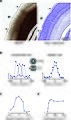 The middle temporal area (MT) of marmoset.jpg 983 × 1,661; 1,021 KB
-
 Visual field maps.jpg 886 × 886; 106 KB
-
 Wandell maps.jpg 1,181 × 886; 114 KB
-
 Wat waar.PNG 616 × 443; 113 KB


_of_marmoset.jpg)


