Category:Mikael Häggström/Micrographs of blood
Jump to navigation
Jump to search

These images were created by Mikael Häggström, M.D.
- User info
- Reusing images
Media in category "Mikael Häggström/Micrographs of blood"
The following 19 files are in this category, out of 19 total.
-
 Blood smear of Babesia microti, annotated.png 1,697 × 1,689; 2.02 MB
-
 Blood smear of Babesia microti, original.png 1,989 × 1,641; 2.63 MB
-
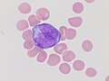
 Cytology of precursor (blast) cell.png 1,291 × 903; 1.32 MB
-
 KB stain original.jpg 2,048 × 1,532; 282 KB
-
 KB stain, annotated.jpg 1,195 × 873; 130 KB
-

-
 Micrograph of an echinocyte on a peripheral blood smear.jpg 216 × 224; 17 KB
-
 Micrograph of an elliptocyte on a peripheral blood smear 02.jpg 217 × 199; 11 KB
-
 Pap smear of a monocyte with nuclear smearing artifact.jpg 875 × 399; 79 KB
-

-
-

-

-

-

-
 Peripheral blood smear of acute promyelocytic leukemia, hypogranular variant.png 2,405 × 1,953; 2.34 MB
-
 Peripheral blood smear with polychromasia.jpg 290 × 240; 17 KB
-
 Poikilocytes - Red blood cell types.jpg 925 × 1,107; 200 KB
-
 Smudge cell in a peripheral blood smear.jpg 509 × 441; 43 KB


_cell.png)


.jpg)
.jpg)
.jpg)
.jpg)
.jpg)
.jpg)


