Category:Microscopic images of Bacillus
Jump to navigation
Jump to search
Subcategories
This category has only the following subcategory.
Media in category "Microscopic images of Bacillus"
The following 62 files are in this category, out of 62 total.
-
 20101017 175758 Bacilli.jpg 3,072 × 2,304; 290 KB
-
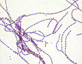 Anthrax cells.jpg 1,500 × 1,175; 156 KB
-
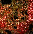
 Anthrax color enhanced micrograph.JPG 1,636 × 1,279; 661 KB
-
 B. subtilis.jpg 1,688 × 1,752; 2.12 MB
-
 Bacilli and cocci.jpg 500 × 500; 143 KB
-
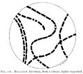 Bacilli of Anthrax from a culture Wellcome M0015725.jpg 3,492 × 3,134; 552 KB
-
 Bacillus anthracis Gram.jpg 2,892 × 1,940; 6.46 MB
-
 Bacillus anthracis hiss.jpg 444 × 448; 52 KB
-
 Bacillus anthracis Indian Ink capsule stain.tif 2,803 × 2,234; 17.92 MB
-
 Bacillus anthracis PHIL 1792.tif 2,545 × 2,332; 16.99 MB
-
 Bacillus anthracis Wellcome M0014322.jpg 3,394 × 3,213; 4.95 MB
-
 Bacillus anthracis.jpg 328 × 246; 53 KB
-
 Bacillus anthracis.png 2,545 × 2,332; 4.53 MB
-
 Bacillus cereus (15469201572).jpg 1,756 × 988; 2.47 MB
-
 Bacillus cereus and Escherichia coli.jpg 3,264 × 2,448; 1.67 MB
-
 Bacillus cereus endospore stain.jpg 1,280 × 1,024; 76 KB
-
 Bacillus cereus Gram.jpg 600 × 450; 17 KB
-
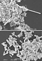
 Bacillus cereus SEM-cr.jpg 2,428 × 1,600; 895 KB
-
 Bacillus cereus SEM.jpg 4,802 × 2,016; 1.65 MB
-
 Bacillus clausii Enterogermina.png 4,000 × 3,200; 9.53 MB
-
 Bacillus coagulans 01.jpg 700 × 460; 56 KB
-
 Bacillus megaterium 1000x magnification.png 1,397 × 1,047; 2.51 MB
-
 Bacillus megaterium DSM-90 cells.jpg 1,546 × 1,138; 154 KB
-
 Bacillus odysseyi.jpg 503 × 309; 26 KB
-
 Bacillus simplex (1657741).jpg 2,048 × 1,532; 177 KB
-
 Bacillus species.jpg 2,080 × 1,536; 2.36 MB
-
 Bacillus subtilis (2).jpg 478 × 318; 90 KB
-
 Bacillus subtilis endospore stain.png 2,119 × 2,119; 6.74 MB
-
 Bacillus subtilis gram stain CDC PHIL 19261.jpg 700 × 460; 19 KB
-
 Bacillus subtilis Gram stain.jpg 2,080 × 1,536; 2.31 MB
-
 Bacillus subtilis Gram.jpg 500 × 375; 38 KB
-
 Bacillus subtilis image.jpg 1,224 × 1,632; 545 KB
-
 Bacillus subtilis R0179.jpg 457 × 552; 59 KB
-
 Bacillus subtilis Spore.jpg 500 × 375; 34 KB
-
 Bacillus subtilis stain CDC PHIL 19260.jpg 700 × 460; 50 KB
-
 Bacillus subtilis stained with Nile Red.jpg 797 × 797; 153 KB
-
 Bacillus subtilis.jpg 1,376 × 1,032; 73 KB
-
 Bacillus Subtilis.jpg 150 × 150; 10 KB
-
 Bacillus thuringiensis-S.jpg 1,698 × 1,619; 678 KB
-
 Bacillus thuringiensis.jpg 2,080 × 1,536; 1.99 MB
-
 BacillusCereus.jpg 585 × 481; 35 KB
-
 Biology laboratory work, bacteria- bacillus subtilies.JPG 2,048 × 2,048; 629 KB
-
 Bt 4A4 (24866589842).jpg 2,448 × 3,264; 7.06 MB
-
 Comparison of Room Temperature and Cryo Methods (8531613420).jpg 300 × 418; 59 KB
-
 Eubacteria (259 10) Bacillus subtilis bacteria.jpg 3,751 × 2,401; 1.97 MB
-
 Eubacteria (259 11) Bacillus subtilis bacteria.jpg 3,751 × 2,401; 2.03 MB
-
 Eubacteria (26 2 87) Bacillus subtilis; spores stained green.jpg 2,776 × 1,777; 1.45 MB
-
 Filamentation 1.jpg 1,126 × 802; 159 KB
-
 Filamentation 2.jpg 1,127 × 1,615; 274 KB
-
 Gram positive bacilli.jpg 2,068 × 1,336; 1.48 MB
-
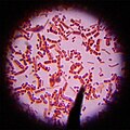
 Gram Stain Anthrax.jpg 600 × 405; 41 KB
-
 Live Sample of Bacillus Cereus Through a 10x Magnification Light Microscope Lens.jpg 3,024 × 4,032; 1.22 MB
-
 Phase contrast image of L-form bacteria-Mark Leaver Newcastle University.jpeg 1,040 × 787; 454 KB
-
 Phase contrast image of L-form bacteria-Mark Leaver Newcastle University.tif 1,040 × 787; 1.59 MB
-
 Spores of Bacillus subtilis.tif 1,033 × 896; 1.31 MB
-
 Stained Bacillus subtilis.jpg 503 × 671; 53 KB
-
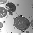 TEM of L-form bacteria-Mark Leaver Newcastle University.jpeg 1,824 × 1,928; 1.73 MB
-
 TEM of L-form bacteria-Mark Leaver Newcastle University.tif 1,824 × 1,928; 6.73 MB
-
 Wide field EM of L-form bacteria-Mark Leaver Newcastle University.jpeg 1,824 × 1,940; 1.98 MB
-
 Wide field EM of L-form bacteria-Mark Leaver Newcastle University.tif 1,824 × 1,940; 6.78 MB
-
 Микроколония Bacillus subtilis, т.п. х400 (2).jpg 1,292 × 968; 163 KB
-
 Сенная палочка.jpg 678 × 678; 87 KB


.jpg)


.jpg)

.jpg)


.jpg)
.jpg)
_Bacillus_subtilis_bacteria.jpg)
_Bacillus_subtilis_bacteria.jpg)
_Bacillus_subtilis;_spores_stained_green.jpg)


.jpg)
