Category:Histopathology of urinary bladder
Jump to navigation
Jump to search
Radiology: Ultrasound · X-ray · Computed tomography · Magnetic resonance · Positron emission tomography | Anatomical pathology: Gross pathology · Histopathology | Other: Cystoscopy · Epidemiology (Map → World) | File format: SVG | 
Subcategories
This category has the following 4 subcategories, out of 4 total.
C
N
Media in category "Histopathology of urinary bladder"
The following 83 files are in this category, out of 83 total.
-
 BCG-induced granulomatous cystitis, high mag.jpg 4,272 × 2,848; 6.19 MB
-
 BCG-induced granulomatous cystitis, intermed. mag.jpg 4,272 × 2,848; 7.66 MB
-
 BCG-induced granulomatous cystitis, low mag.jpg 4,272 × 2,848; 8.42 MB
-
 BCG-induced granulomatous cystitis, very high mag.jpg 4,272 × 2,848; 4.92 MB
-
 Bladder neck invasion in prostate cancer -- very low mag.jpg 6,000 × 4,000; 9.79 MB
-
 Bladder with benign large mesenchymal cells - alt -- high mag.jpg 4,000 × 6,000; 16.91 MB
-
 Bladder with benign large mesenchymal cells - alt -- intermed mag.jpg 4,000 × 6,000; 13.99 MB
-
 Bladder with benign large mesenchymal cells - alt -- very high mag.jpg 4,000 × 6,000; 12.01 MB
-
 Bladder with benign large mesenchymal cells -- high mag.jpg 4,000 × 6,000; 10.42 MB
-
 Bladder with benign large mesenchymal cells -- intermed mag.jpg 4,000 × 6,000; 14.25 MB
-
 Bladder with benign large mesenchymal cells -- low mag.jpg 6,000 × 4,000; 10.76 MB
-
 Bladder with benign large mesenchymal cells -- very high mag.jpg 4,000 × 6,000; 12.16 MB
-
 Bladder with benign large mesenchymal cells -- very low mag.jpg 6,000 × 4,000; 10.75 MB
-
 Bullous cystitis, intermed. mag.jpg 4,272 × 2,848; 3.89 MB
-
 Bullous cystitis, low mag.jpg 4,272 × 2,848; 4.24 MB
-
 Cystitis cystica, high mag.jpg 4,272 × 2,848; 6.04 MB
-
 Cystitis cystica, intermed. mag.jpg 4,272 × 2,848; 6.94 MB
-
 Cystitis cystica, low mag.jpg 4,272 × 2,848; 6.77 MB
-
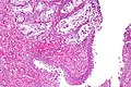
 Cystitis glandularis intestinal type, high mag.jpg 4,272 × 2,848; 1.67 MB
-
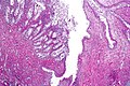
 Cystitis glandularis intestinal type, intermed. mag.jpg 4,272 × 2,848; 2.12 MB
-
 Cystitis glandularis intestinal type, low mag.jpg 4,272 × 2,848; 6.7 MB
-
 Cystitis glandularis intestinal type, very high mag.jpg 4,272 × 2,848; 1.36 MB
-
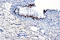 Cystitis glandularis usual type - CDX2, high mag.jpg 4,272 × 2,848; 1.13 MB
-
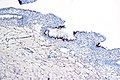 Cystitis glandularis usual type - CDX2, intermed. mag.jpg 4,272 × 2,848; 1.18 MB
-
 Cystitis glandularis usual type - CDX2, very high mag.jpg 4,272 × 2,848; 1,024 KB
-
 Eosinophilic cystitis, high mag.1.jpg 4,272 × 2,848; 5.91 MB
-
 Eosinophilic cystitis, high mag.2.jpg 4,272 × 2,848; 6.41 MB
-
 Eosinophilic cystitis, intermed. mag.jpg 4,272 × 2,848; 7.1 MB
-
 Eosinophilic cystitis, low mag.jpg 4,272 × 2,848; 6.05 MB
-
 Eosinophilic cystitis, very high mag.1.jpg 4,272 × 2,848; 5.02 MB
-
 Eosinophilic cystitis, very high mag.2.jpg 4,272 × 2,848; 5.46 MB
-
 Exophytic urothelial papilloma, intermed. mag.jpg 4,272 × 2,848; 4.03 MB
-
 Exophytic urothelial papilloma, low mag.jpg 4,272 × 2,848; 5.54 MB
-
 Flat urothelial hyperplasia, high mag.jpg 4,272 × 2,848; 3.72 MB
-
 Flat urothelial hyperplasia, intermed. mag.jpg 4,272 × 2,848; 3.36 MB
-
 Flat urothelial hyperplasia, low mag.jpg 4,272 × 2,848; 3.98 MB
-
 Flat urothelial hyperplasia, very high mag.jpg 4,272 × 2,848; 3.95 MB
-
 Follicular cystitis, high mag.jpg 4,272 × 2,848; 5.98 MB
-
 Follicular cystitis, intermed. mag.jpg 4,272 × 2,848; 6.45 MB
-
 Follicular cystitis, low mag.jpg 4,272 × 2,848; 5.97 MB
-
 Granulomatous inflammation of bladder neck.jpg 4,272 × 2,848; 5.08 MB
-
 HK hemorr, inf and the other.tif 4,080 × 3,072; 35.88 MB
-
 Inverted urothelial papilloma, high mag.1.jpg 4,272 × 2,848; 1.6 MB
-
 Inverted urothelial papilloma, high mag.2.jpg 4,272 × 2,848; 5.1 MB
-
 Inverted urothelial papilloma, intermed. mag.1.jpg 4,272 × 2,848; 2.48 MB
-
 Inverted urothelial papilloma, intermed. mag.2.jpg 4,272 × 2,848; 7.1 MB
-
 Inverted urothelial papilloma, low mag.jpg 4,272 × 2,848; 4.91 MB
-
 Inverted urothelial papilloma, very high mag.jpg 4,272 × 2,848; 5 MB
-
 Keratinizing squamous metaplasia of the urinary bladder, high mag.1.jpg 4,272 × 2,848; 3.05 MB
-
 Keratinizing squamous metaplasia of the urinary bladder, high mag.2.jpg 4,272 × 2,848; 1.47 MB
-
 Keratinizing squamous metaplasia of the urinary bladder, intermed. mag.jpg 4,272 × 2,848; 3.49 MB
-
 Keratinizing squamous metaplasia of the urinary bladder, low mag.jpg 4,272 × 2,848; 1.93 MB
-
 Malakoplakia of the urinary bladder, high mag.jpg 4,272 × 2,848; 5.54 MB
-
 Malakoplakia of the urinary bladder, intermed. mag.jpg 4,272 × 2,848; 6.98 MB
-
 Malakoplakia of the urinary bladder, low mag.jpg 4,272 × 2,848; 4.99 MB
-
 Malakoplakia of the urinary bladder, very high mag.jpg 4,272 × 2,848; 4.82 MB
-
 Nonkeratinizing & keratinizing squamous metaplasia of the urinary bladder, high mag.jpg 4,272 × 2,848; 3.66 MB
-
 Nonkeratinizing & keratinizing squamous metaplasia of the urinary bladder, intermed. mag.jpg 4,272 × 2,848; 4.33 MB
-
 Nonkeratinizing & keratinizing squamous metaplasia of the urinary bladder, low mag.jpg 4,272 × 2,848; 4.27 MB
-
 Nonkeratinizing squamous metaplasia of the urinary bladder, high mag.jpg 4,272 × 2,848; 3.42 MB
-
 Papillary cystitis, high mag.jpg 4,272 × 2,848; 1.28 MB
-
 Papillary cystitis, intermed. mag.jpg 4,272 × 2,848; 1.8 MB
-
 Papillary cystitis, low mag.jpg 4,272 × 2,848; 1.73 MB
-
 Polypoid cystitis, high mag.jpg 4,272 × 2,848; 1.74 MB
-
 Polypoid cystitis, intermed. mag.jpg 4,272 × 2,848; 2.07 MB
-
 Polypoid cystitis, low mag.jpg 4,272 × 2,848; 1.78 MB
-
 Transactions of the Southern Surgical and Gynecological Association (1910) (14745714136).jpg 3,040 × 1,870; 1.73 MB
-
 Urinary bladder - biopsy site reaction, high mag.jpg 4,272 × 2,848; 6.46 MB
-
 Urinary bladder - biopsy site reaction, intermed. mag.jpg 4,272 × 2,848; 7.86 MB
-
 Urinary bladder - biopsy site reaction, low mag.jpg 4,272 × 2,848; 6.82 MB
-
 Urinary bladder - biopsy site reaction, very high mag.jpg 4,272 × 2,848; 4.93 MB
-
 Urinary bladder with glycogenated squamous epithelium, high mag.jpg 4,272 × 2,848; 1.74 MB
-
 Urinary bladder with glycogenated squamous epithelium, intermed. mag.jpg 4,272 × 2,848; 2.34 MB
-
 Urinary bladder with glycogenated squamous epithelium, low mag.jpg 4,272 × 2,848; 1.56 MB
-
 Urinary bladder with glycogenated squamous epithelium, very high mag.jpg 4,272 × 2,848; 4.98 MB
-
 Urothelial hyperplasia, high mag.jpg 4,272 × 2,848; 4.64 MB
-
 Urothelial hyperplasia, intermed. mag.jpg 4,272 × 2,848; 3.84 MB
-
 Urothelial hyperplasia, very high mag.jpg 4,272 × 2,848; 4.76 MB
-
 Urothelial papilloma, high mag.1.jpg 4,272 × 2,848; 2.6 MB
-
 Urothelial papilloma, high mag.2.jpg 4,272 × 2,848; 5.19 MB
-
 Urothelial papilloma, intermed. mag.1.jpg 4,272 × 2,848; 3.54 MB
-
 Urothelial papilloma, intermed. mag.2.jpg 4,272 × 2,848; 4.95 MB
-
 Urothelial papilloma, low mag.jpg 4,272 × 2,848; 4.66 MB


_(14745714136).jpg)