Category:Histopathology of the breast
Jump to navigation
Jump to search
Wikimedia category | |||||
| Upload media | |||||
| Instance of | |||||
|---|---|---|---|---|---|
| |||||
Subcategories
This category has the following 11 subcategories, out of 11 total.
A
G
H
N
T
Pages in category "Histopathology of the breast"
This category contains only the following page.
Media in category "Histopathology of the breast"
The following 42 files are in this category, out of 42 total.
-
 Apocrine metaplasia - alt -- very high mag.jpg 4,272 × 2,848; 4.18 MB
-
 Apocrine metaplasia -- high mag.jpg 4,272 × 2,848; 4.8 MB
-
 Apocrine metaplasia -- intermed mag.jpg 4,272 × 2,848; 5.57 MB
-
 Apocrine metaplasia -- low mag.jpg 4,272 × 2,848; 5.85 MB
-
 Apocrine metaplasia -- very high mag.jpg 4,272 × 2,848; 4.1 MB
-
 Atypical lobular hyperplasia - E-cadherin -- high mag.jpg 6,000 × 4,000; 15.06 MB
-
 Atypical lobular hyperplasia - E-cadherin -- intermed mag.jpg 6,000 × 4,000; 12.61 MB
-
 Atypical lobular hyperplasia -- high mag.jpg 6,000 × 4,000; 15.72 MB
-
 Atypical lobular hyperplasia -- intermed mag.jpg 6,000 × 4,000; 15.02 MB
-
 Atypical lobular hyperplasia -- low mag.jpg 6,000 × 4,000; 12.34 MB
-
 Benign breast - E-cadherin - alt -- high mag.jpg 6,000 × 4,000; 12.51 MB
-
 Benign breast - E-cadherin -- high mag.jpg 6,000 × 4,000; 11.88 MB
-
 Benign breast - E-cadherin -- intermed mag.jpg 6,000 × 4,000; 10.8 MB
-
 Breast tissue showing fat necrosis 4X.jpg 1,447 × 1,132; 198 KB
-
 Breast tissue showing fat necrosis.jpg 997 × 749; 101 KB
-
 Calcifications of benign breast glands.jpg 2,048 × 1,532; 754 KB
-
 Columnar cell change -- high mag.jpg 6,000 × 4,000; 14.02 MB
-
 Columnar cell change -- intermed mag.jpg 6,000 × 4,000; 15.58 MB
-
 Columnar cell change -- very high mag.jpg 6,000 × 4,000; 13.7 MB
-
 Gynecomastoid hyperplasia - high mag.jpg 4,272 × 2,848; 4.78 MB
-
 Gynecomastoid hyperplasia - intermed mag.jpg 4,272 × 2,848; 5.08 MB
-
 Gynecomastoid hyperplasia - low mag.jpg 4,272 × 2,848; 5.07 MB
-
 Gynecomastoid hyperplasia - very high mag.jpg 4,272 × 2,848; 3.72 MB
-
 Gynecomastoid hyperplasia -2- high mag.jpg 4,272 × 2,848; 4.73 MB
-
 Gynecomastoid hyperplasia -2- intermed mag.jpg 4,272 × 2,848; 5.22 MB
-
 Gynecomastoid hyperplasia -2- low mag.jpg 4,272 × 2,848; 5.2 MB
-
 Gynecomastoid hyperplasia -2- very high mag.jpg 4,272 × 2,848; 3.93 MB
-
 Hibridación in situ fluorescente (FISH) del Gen Her2 (no amplificado) (mama) (05).jpg 1,920 × 1,036; 239 KB
-
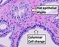 Histopathology of flat epithelial atypia and columnar cell change.jpg 1,015 × 805; 244 KB
-
 Histopathology of stromal fibrosis of the breast.jpg 519 × 548; 78 KB
-
 IBC-HE.png 2,592 × 1,944; 9.1 MB
-
 Lactational change - high mag.jpg 4,272 × 2,848; 6.33 MB
-
 Lactational change - intermed mag.jpg 4,272 × 2,848; 5.8 MB
-
 Lactational change - low mag.jpg 4,272 × 2,848; 5.99 MB
-
 Lactational change - very high mag.jpg 4,272 × 2,848; 4.54 MB
-
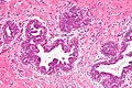
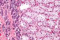
 Proliferative fibrocystic changes -- high mag.jpg 4,272 × 2,848; 4.87 MB
-
 Proliferative fibrocystic changes -- intermed mag.jpg 4,272 × 2,848; 5.06 MB
-
 Proliferative fibrocystic changes -- low mag.jpg 4,272 × 2,848; 5.4 MB
-
 Proliferative fibrocystic changes -- very high mag.jpg 4,272 × 2,848; 4.07 MB
-
 Pseudoangiomatous stromal hyperplasia - very high mag.jpg 4,272 × 2,848; 3.97 MB
-
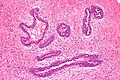
 Radial scar of breast - intermed mag.jpg 6,000 × 4,000; 14.27 MB
-
 Radial scar of breast - low mag.jpg 6,000 × 4,000; 13.38 MB


_del_Gen_Her2_(no_amplificado)_(mama)_(05).jpg)


