Category:CT images of congenital anomalies of the atlas and the cervico-occipital transition
Jump to navigation
Jump to search
| 
Media in category "CT images of congenital anomalies of the atlas and the cervico-occipital transition"
The following 19 files are in this category, out of 19 total.
-
 Akzessorischer Knochen am Unterrand des vorderen Atlasbogens 26W - CT - 001.jpg 1,944 × 968; 130 KB
-
 Akzessorisches Ossikel am vorderen Atlasbogen - CT sag und ax 001.jpg 1,286 × 980; 111 KB
-
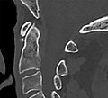
 Akzessorisches Ossikel am vorderen Atlasbogen - CT sag und cor 001.jpg 1,286 × 980; 115 KB
-
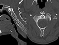
 Akzessorisches Ossikel am vorderen Atlasbogen 92M - CT sagittal - 001.jpg 1,117 × 1,013; 71 KB
-
 Atlantookzipitale und atlantoaxiale Fusion 87W - CT - 001.jpg 2,261 × 873; 231 KB
-
 Atlasbogenspalt - 001.jpg 1,158 × 1,401; 77 KB
-
 Atlasbogenspalt dorsal 83W - CR seitlich und CT Volumen Rendering - 001.jpg 1,591 × 867; 263 KB
-
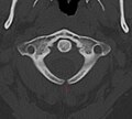
 Atlasbogenspalt hinten und Sklerosierung vorne - CT axial - 001.jpg 1,280 × 920; 78 KB
-
 Atlasbogenspalt.jpg 1,142 × 1,031; 110 KB
-

-
 Bogenschlussstoerung des Atlas 92M - CT axial und sagittal - 001.jpg 1,521 × 835; 121 KB
-
 Dorsale Bogenschlussstoerung am Atlas 72M - CT axial - 001.jpg 835 × 1,105; 82 KB
-
 Hinterer Atlasbogen-Spalt 33W - CT axial und Volumen Rendering - 001.jpg 1,534 × 676; 173 KB
-
 Os epitransversarium links 44W - CT und CR - 001 - Annotation.jpg 2,891 × 1,036; 326 KB
-
 Os epitransversarium links 44W - CT und CR - 001.jpg 2,891 × 1,036; 322 KB
-

-
 Processus paracondylicus links 87W - CT cor und VR - 001 - Annotation.jpg 1,575 × 810; 182 KB
-
 Processus paracondylicus links 87W - CT cor und VR - 001.jpg 1,575 × 810; 181 KB
-
 Split Atlas 50jw - CT ax und Volumen - 001.png 1,622 × 765; 1.33 MB


