Category:Microscopic images of Arthropoda
Jump to navigation
Jump to search
Subcategories
This category has the following 5 subcategories, out of 5 total.
*
A
C
I
M
Media in category "Microscopic images of Arthropoda"
The following 42 files are in this category, out of 42 total.
-
 Acari (265 02) Total preparation.jpg 3,751 × 2,401; 1.26 MB
-
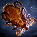
 Big tick.jpg 2,048 × 1,536; 179 KB
-
 Castor bean tick (248 14) Total preparation - larva and egg.jpg 3,751 × 2,400; 1.12 MB
-
 Castor bean tick (250 16) Adult.jpg 3,748 × 2,399; 1.38 MB
-
 Castor bean tick (265 28) Total preparation - larva and part of an egg.jpg 3,750 × 2,400; 1.05 MB
-
 Castor bean tick (265 29) Adult head.jpg 3,750 × 2,400; 1.3 MB
-
 Centipede on microscope.jpg 1,080 × 1,080; 80 KB
-
 Collembola sp. (1).jpg 2,000 × 2,000; 2.64 MB
-
 Collembola sp. (2).jpg 2,400 × 2,400; 4.22 MB
-
 Collembolla Symphipleona.jpg 440 × 213; 10 KB
-
 Difference between Glomerida (Myriapoda) and Isopoda (Crustacea).jpg 2,943 × 3,042; 799 KB
-
 Flea microphotograph - By Arthur E. Durham.jpg 1,760 × 2,000; 153 KB
-
 Haarling Meerschweinchen.jpg 403 × 403; 24 KB
-
 Ixodes ricinus DF.jpg 1,185 × 1,196; 965 KB
-
 Ixodes ricinus IMG 4828.JPG 1,113 × 1,723; 359 KB
-
 Ixodes ricinus IMG 4829.JPG 2,228 × 2,412; 713 KB
-
 Ixodes ricinus IMG 4846.JPG 1,441 × 2,336; 659 KB
-
 Mikrofoto.de-Kugelmilbe.jpg 1,000 × 667; 99 KB
-
 Muscle structure of forcipule of a giant Australian centipede.tif 2,778 × 1,338; 17.08 MB
-
 Parasite150059-fig1 - Chewing lice of genus Ricinus.tif 2,343 × 1,931; 9.12 MB
-
 Parasite150059-fig1 - Ricinus vaderi.tif 795 × 1,901; 3.15 MB
-
 Pengamatan Mikroskopis Arthropoda Air.jpg 4,741 × 4,741; 3.17 MB
-
 Phase-contrast x-ray image of spider.jpg 682 × 509; 241 KB
-
 Polyplax spinulosa louse under microscope.jpg 192 × 168; 22 KB
-
 Protura (Acerentomon species) micrograph.jpg 504 × 256; 143 KB
-
 Protura specimen (Acerentomon species) micrograph.jpg 576 × 411; 241 KB
-
 Protura specimen.jpg 786 × 570; 149 KB
-
Psoroptes on a rabbit ear crust.webm 1 min 1 s, 1,280 × 720; 8.31 MB
-
 Psoroptes-life-cycle-diagram.png 383 × 351; 57 KB
-
 Raubmilbe Detail 1.jpg 709 × 595; 266 KB
-
 Sanja.jpg 640 × 600; 238 KB
-
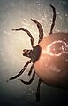
 Small tick.jpg 2,048 × 1,536; 1.9 MB
-
 Spider leg (250 08) Spider leg (Arachnida).jpg 3,750 × 2,400; 1.83 MB
-
 Stigma of arachnid trachea (250 03) Stigma of arachnid trachea.jpg 3,751 × 2,401; 1.19 MB
-
 Stinde Mikrofoto2.jpg 207 × 273; 64 KB
-
 The expression of C12orf29 in forcipule muscle in giant Australian centipede.jpg 2,131 × 1,014; 258 KB
-
 Tick Salivary Gland Infected with Langat Virus (47974625372).jpg 2,048 × 2,048; 4.07 MB
-
 Ácaro hallado en alimento de peces.jpg 1,800 × 2,620; 1.83 MB
-
 Мелкие членистоногие под микроскопом.jpg 5,120 × 3,200; 4.83 MB
-
 Паутинный клещ Tetranychus urticae.png 3,786 × 1,398; 6.31 MB
-
 Последний выдох.jpg 1,924 × 2,579; 5.5 MB
-
 Почвенный клещ.jpg 7,000 × 10,501; 11.3 MB
_Total_preparation.jpg)

_Total_preparation_-_larva_and_egg.jpg)
_Adult.jpg)
_Total_preparation_-_larva_and_part_of_an_egg.jpg)
_Adult_head.jpg)

.jpg)
.jpg)

_and_Isopoda_(Crustacea).jpg)


_micrograph.jpg)
_micrograph.jpg)


_Spider_leg_(Arachnida).jpg)
_Stigma_of_arachnid_trachea.jpg)


.jpg)


