File:Schematic of cortical areas involved with pain processing and fMRI cropped.jpg
Jump to navigation
Jump to search

Size of this preview: 800 × 541 pixels. Other resolutions: 320 × 216 pixels | 640 × 432 pixels | 1,024 × 692 pixels | 1,172 × 792 pixels.
Original file (1,172 × 792 pixels, file size: 211 KB, MIME type: image/jpeg)
Captions
Captions
Add a one-line explanation of what this file represents
| Description |
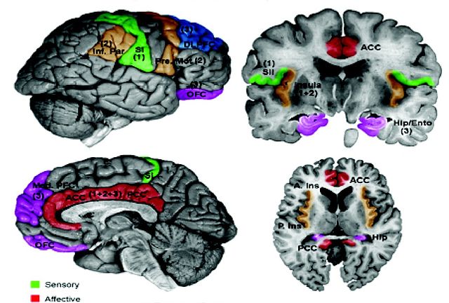
English: Examples of CNS Functional Measures. A. Schematic of cortical areas involved with pain processing. The highlighted areas summarize areas found active in previous functional imaging studies. Color-coding reflects the hypothesized role of each area in processing the different psychological dimensions of pain. Numbers in parentheses indicate the relative involvement of these areas during different temporal stages of the pain experience. Areas displayed include insula, anterior cingulate cortex (ACC), posterior cingulate cortex (PCC), primary somatosensory cortex (SI), secondary somatosensory cortex (SII), inferior parietal lobe (Inf. Par), dorsolateral prefrontal cortex (DLPFC), pre-motor cortex (Pre-Mot), orbitofrontal cortex (OFC), medial prefrontal cortex (Med. PFC), posterior insula (P. Ins), anterior insula (A. Ins), hippocampus (Hip), entorhinal cortex (Ento). [Reprinted with permission from Casey and Tran, 2006]. For examples of brainstem involvement in pain processing, please refer to Tracey and Iannetti ([52]). B. Example of fMRI responses to painful phasic thermal stimulation to the forehead in a cohort of 12 subjects. (Moulton et al., unpublished observations). Borsook et al. Molecular Pain 2007 3:25 doi:10.1186/1744-8069-3-25 |
| Date | (UTC) |
| Source | |
| Author |
|
| This is a retouched picture, which means that it has been digitally altered from its original version. Modifications: Cropped. The original can be viewed here: Schematic of cortical areas involved with pain processing and fMRI.jpg:
|
I, the copyright holder of this work, hereby publish it under the following license:
This file is licensed under the Creative Commons Attribution 2.0 Generic license.
- You are free:
- to share – to copy, distribute and transmit the work
- to remix – to adapt the work
- Under the following conditions:
- attribution – You must give appropriate credit, provide a link to the license, and indicate if changes were made. You may do so in any reasonable manner, but not in any way that suggests the licensor endorses you or your use.
Original upload log[edit]
This image is a derivative work of the following images:
- File:Schematic_of_cortical_areas_involved_with_pain_processing_and_fMRI.jpg licensed with Cc-by-2.0
- 2009-08-27T15:01:26Z CopperKettle 1200x1383 (386847 Bytes) {{Information |Description={{en|1=Examples of CNS Functional Measures. A. Schematic of cortical areas involved with pain processing. The highlighted areas summarize areas found active in previous functional imaging studies. C
Uploaded with derivativeFX
File history
Click on a date/time to view the file as it appeared at that time.
| Date/Time | Thumbnail | Dimensions | User | Comment | |
|---|---|---|---|---|---|
| current | 13:39, 5 June 2010 | | 1,172 × 792 (211 KB) | Anthonyhcole (talk | contribs) | {{Information |Description={{en|1=Examples of CNS Functional Measures. A. Schematic of cortical areas involved with pain processing. The highlighted areas summarize areas found active in previous functional imaging studies. Color-coding reflects the hypot |
You cannot overwrite this file.
File usage on Commons
There are no pages that use this file.
File usage on other wikis
The following other wikis use this file:
- Usage on en.wikipedia.org
- Usage on en.wikiquote.org
- Usage on en.wikiversity.org
- Usage on es.wikipedia.org
- Usage on ga.wikipedia.org
- Usage on it.wikipedia.org
- Usage on ru.wikiversity.org