File:Schematic diagram of the human eye.png
Schematic_diagram_of_the_human_eye.png (600 × 550 pixels, file size: 54 KB, MIME type: image/png)
Captions
Captions
Summary[edit]
| Description |
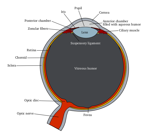
English: Schematic diagram of the human eye
|
|||||
| Source |
Own work using: |
|||||
| Author | Delta G | |||||
| Other versions |
[] All eye
By languages
For translate
Anterior segment
|


Licensing[edit]
| This file is licensed under the Creative Commons Attribution-Share Alike 3.0 Unported license. Subject to disclaimers. | ||
| ||
| This licensing tag was added to this file as part of the GFDL licensing update. |

|
Permission is granted to copy, distribute and/or modify this document under the terms of the GNU Free Documentation License, Version 1.2 or any later version published by the Free Software Foundation; with no Invariant Sections, no Front-Cover Texts, and no Back-Cover Texts. A copy of the license is included in the section entitled GNU Free Documentation License. Subject to disclaimers. |
Eye Anatomy[edit]
A guide to the many parts of the human eye and how they function.
The ability to see is dependent on the actions of several structures in and around the eyeball. The graphic below lists many of the essential components of the eye's optical system.
When you look at an object, light rays are reflected from the object to the cornea, which is where the miracle begins. The light rays are bent, refracted and focused by the cornea, lens, and vitreous. The lens' job is to make sure the rays come to a sharp focus on the retina. The resulting image on the retina is upside-down. Here at the retina, the light rays are converted to electrical impulses which are then transmitted through the optic nerve, to the brain, where the image is translated and perceived in an upright position!
Think of the eye as a camera. A camera needs a lens and a film to produce an image. In the same way, the eyeball needs a lens (cornea, crystalline lens, vitreous) to refract, or focus the light and a film (retina) on which to focus the rays. If any one or more of these components is not functioning correctly, the result is a poor picture. The retina represents the film in our camera. It captures the image and sends it to the brain to be developed. The macula is the highly sensitive area of the retina. The macula is responsible for our critical focusing vision. It is the part of the retina most used. We use our macula to read or to stare intently at an object.
File history
Click on a date/time to view the file as it appeared at that time.
| Date/Time | Thumbnail | Dimensions | User | Comment | |
|---|---|---|---|---|---|
| current | 12:00, 5 June 2006 | | 600 × 550 (54 KB) | Eliashc (talk | contribs) | optimized using optipng. |
| 05:37, 14 March 2005 |  | 600 × 550 (73 KB) | Delta G (talk | contribs) | Schematic diagram of the human eye(zonule fibers -> zonular fibers(more common)) | |
| 04:58, 14 March 2005 |  | 600 × 550 (72 KB) | Delta G (talk | contribs) | Schematic diagram of the human eye(smaller, vitreous fluid -> vitreous humor) | |
| 04:36, 14 March 2005 |  | 866 × 793 (115 KB) | Delta G (talk | contribs) | Schematic diagram of the human eye |
You cannot overwrite this file.
File usage on Commons
There are no pages that use this file.
File usage on other wikis
The following other wikis use this file:
- Usage on ar.wikipedia.org
- Usage on en.wikipedia.org
- Usage on fa.wikipedia.org