File:Sacroiliitis MRI ar1934-5.gif
Jump to navigation
Jump to search

Size of this preview: 379 × 599 pixels. Other resolutions: 152 × 240 pixels | 304 × 480 pixels | 1,004 × 1,586 pixels.
Original file (1,004 × 1,586 pixels, file size: 1.07 MB, MIME type: image/gif)
Captions
Captions
Add a one-line explanation of what this file represents
| Description |
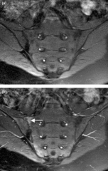
English: Magnetic resonance images of sacroiliac joints: psoriatic arthritis. Shown are T1-weighted semi-coronal magnetic resonance images through the sacroiliac joints (a) before and (b) after intravenous contrast injection. Enhancement is seen at the right sacroiliac joint (arrow), indicating active sacroiliitis. |
| Date | Published: 23 March 2006 |
| Source | Magnetic resonance imaging in psoriatic arthritis: a review of the literature. Arthritis Research & Therapy 2006, 8:207. doi:10.1186/ar1934 |
| Author | Fiona McQueen, Marissa Lassere and Mikkel Østergaard. |
Licensing[edit]
This file is licensed under the Creative Commons Attribution 2.0 Generic license.
- You are free:
- to share – to copy, distribute and transmit the work
- to remix – to adapt the work
- Under the following conditions:
- attribution – You must give appropriate credit, provide a link to the license, and indicate if changes were made. You may do so in any reasonable manner, but not in any way that suggests the licensor endorses you or your use.
| Annotations | This image is annotated: View the annotations at Commons |
File history
Click on a date/time to view the file as it appeared at that time.
| Date/Time | Thumbnail | Dimensions | User | Comment | |
|---|---|---|---|---|---|
| current | 19:33, 6 February 2009 | | 1,004 × 1,586 (1.07 MB) | Stevenfruitsmaak (talk | contribs) | {{Information |Description={{en|1=Magnetic resonance images of sacroiliac joints: psoriatic arthritis. Shown are T1-weighted semi-coronal magnetic resonance images through the sacroiliac joints (a) before and (b) after intravenous contrast injection. Enha |
You cannot overwrite this file.
File usage on Commons
The following 2 pages use this file:
File usage on other wikis
The following other wikis use this file:
- Usage on ar.wikipedia.org
- Usage on az.wikipedia.org
- Usage on bs.wikipedia.org
- Usage on ca.wikipedia.org
- Usage on en.wikipedia.org
- Usage on es.wikipedia.org
- Usage on fa.wikipedia.org
- Usage on hi.wikipedia.org
- Usage on hu.wikipedia.org
- Usage on it.wikipedia.org
- Usage on outreach.wikimedia.org
- Usage on ta.wikipedia.org
- Usage on www.wikidata.org
- Usage on zh.wikipedia.org