File:RER Gland MO.png
Jump to navigation
Jump to search
Size of this preview: 798 × 600 pixels. Other resolutions: 319 × 240 pixels | 639 × 480 pixels | 1,019 × 766 pixels.
Original file (1,019 × 766 pixels, file size: 1.02 MB, MIME type: image/png)
Captions
Captions
Add a one-line explanation of what this file represents
Summary[edit]
| Description |
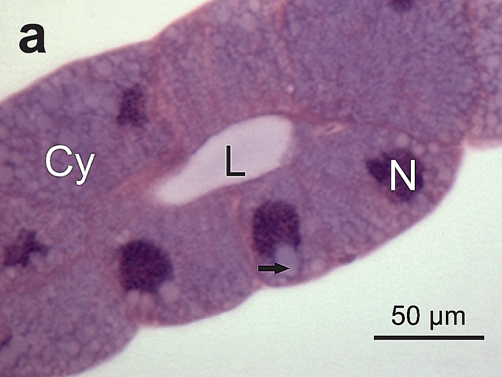
Español: Figura 4. Secciones histológicas de los accesorios de las glándulas salivales de E. heros . a): Epitelio secretor que muestra células cuboideas con núcleo basal (N) y citoplasma con varias vacuolas (flecha). Lumen (L). |
| Date | |
| Source | Ultrastructural analysis of salivary glands in a phytophagous stink bug revealed the presence of unexpected muscles. PLoS ONE 12(6): e0179478. doi:10.1371/journal.pone.0179478 |
| Author | Castellanos N., Martínez L.C., Silva E.H., Teodoro A.V., Serrão J.E., Oliveira E.E. (2017) |
Licensing[edit]
This file is licensed under the Creative Commons Attribution 4.0 International license.
- You are free:
- to share – to copy, distribute and transmit the work
- to remix – to adapt the work
- Under the following conditions:
- attribution – You must give appropriate credit, provide a link to the license, and indicate if changes were made. You may do so in any reasonable manner, but not in any way that suggests the licensor endorses you or your use.
File history
Click on a date/time to view the file as it appeared at that time.
| Date/Time | Thumbnail | Dimensions | User | Comment | |
|---|---|---|---|---|---|
| current | 04:22, 6 July 2021 | | 1,019 × 766 (1.02 MB) | Sanador2.0 (talk | contribs) | Uploaded a work by Castellanos N., Martínez L.C., Silva E.H., Teodoro A.V., Serrão J.E., Oliveira E.E. (2017) from Ultrastructural analysis of salivary glands in a phytophagous stink bug revealed the presence of unexpected muscles. PLoS ONE 12(6): e0179478. doi:10.1371/journal.pone.0179478 with UploadWizard |
You cannot overwrite this file.
File usage on Commons
There are no pages that use this file.
File usage on other wikis
The following other wikis use this file:
- Usage on es.wikipedia.org