File:Mitocondria Axon Presinaptico.PNG
Original file (2,220 × 1,197 pixels, file size: 3.49 MB, MIME type: image/png)
Captions
Captions
Summary[edit]
| Description |
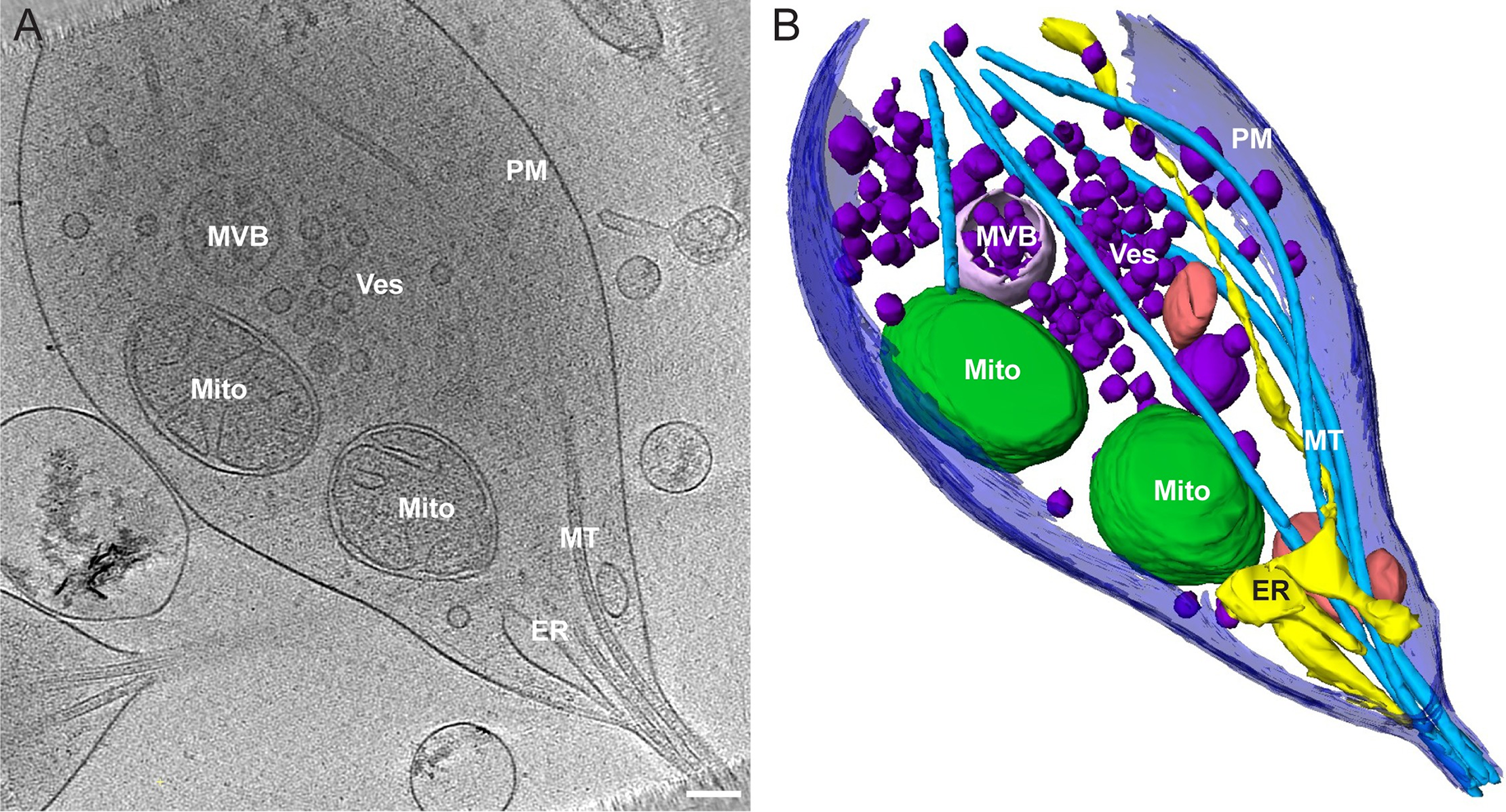
Español: Fig. 2. Reconstrucción tomográfica de una varicosidad presináptica típica y segmento axónico adyacente. (A) Un corte 2D de la reconstrucción tomográfica que muestra la distribución de los orgánulos en la varicosidad (Mito = mitocondria). |
| Date | |
| Source | Morphology of mitochondria in spatially restricted axons revealed by cryo-electron tomography. PLoS Biol 16(9): e2006169 |
| Author | Fischer T.D., Dash P.K., Liu J., Waxham M.N. |
Licensing[edit]
- You are free:
- to share – to copy, distribute and transmit the work
- to remix – to adapt the work
- Under the following conditions:
- attribution – You must give appropriate credit, provide a link to the license, and indicate if changes were made. You may do so in any reasonable manner, but not in any way that suggests the licensor endorses you or your use.
File history
Click on a date/time to view the file as it appeared at that time.
| Date/Time | Thumbnail | Dimensions | User | Comment | |
|---|---|---|---|---|---|
| current | 20:16, 13 May 2020 | | 2,220 × 1,197 (3.49 MB) | Sanador2.0 (talk | contribs) | {{Information |description ={{es|1=Fig. 2. Reconstrucción tomográfica de una varicosidad presináptica típica y segmento axónico adyacente.<br> (A) Un corte 2D de la reconstrucción tomográfica que muestra la distribución de los orgánulos en la varicosidad (Mito = mitocondria).<br> (B) Representación segmentada de todo el volumen del tomograma 3D que se muestra en (A) revelando el tamaño relativo y la distribución espacial del entorno de orgánulos en el segmento de varices y axones. <br> PM M... |
You cannot overwrite this file.
File usage on Commons
There are no pages that use this file.
File usage on other wikis
The following other wikis use this file:
- Usage on es.wikipedia.org