File:Image from page 341 of "Chordate morphology" (1962) (20585824866).jpg

From Wikimedia Commons, the free media repository
Jump to navigation Jump to search

Original file(1,018 × 1,082 pixels, file size: 746 KB, MIME type: image/jpeg)

Captions

Captions

Add a one-line explanation of what this file represents

Summary[edit]

Description

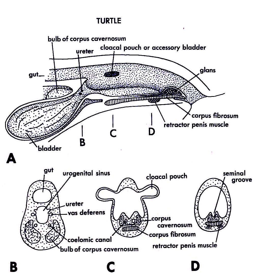
The penis of the turtle, A sgittal section throug cloaca, B, C, D cross sections
Title: Chordate morphology Identifier: chordatemorpholo00joll Year: 1962 (1960s) Authors: Jollie, Malcolm Subjects: Morphology (Animals); Chordata Publisher: New York, Reinhold Contributing Library: MBLWHOI Library Digitizing Sponsor: MBLWHOI Library


View Book Page: Book Viewer About This Book: Catalog Entry View All Images: All Images From Book

Click here to view book online to see this illustration in context in a browseable online version of this book.


Text Appearing Before Image: canal of the mesonephric kidney. From the marginal canal the coiled tubuli mesonephroi lead to the ductus epididy- midis, the anterior end of the nephric duct. The many vari- ations in the testis-kidney connections suggest great plas- ticity of development in this area. The epididymis is well developed and may extend far pos- teriorly before giving way to the slightly convoluted vas de- ferens. The deferent duct joins the ureter of its side, and they open together at the tip of a urogenital papilla. The ducts of either side open through separate papillae. In some reptiles the ureter and vas deferens open separately into the urogenital sinus. The urogenital sinus may be only a pocket of the cloaca lying behind the openings of the anus and the ventral bladder. A vestigial Mullerian duct lies along the body wall lateral to the testis and reproductive tract in general. Two different kinds of intromittent organs are found in reptiles, but Sphenodon lacks such a structure. The lizards and snakes agree in having hemipenes. These are bilateral sacculations of the cloaca (Figure 10-8 B). These sacs ex- tend posteriorly below the skin. When everted, one at a time, they protrude through the cloacal aperture. The distal end is large and rounded, and the stem tapers to a narrow base which attaches at the anterolateral cloacal wall. The structure is grooved to conduct the sperm from the cloacal cavity of the male to that of the female, into which it is everted. The surface of the everted hemipenes is studded with spines. In turtles and crocodilians there is a grooved penis in the floor of the cloaca (Figure 10-38). This contains fibrous and perhaps erectile tissue, a corpus fibrosum. The penis of the turtle is suggestive of that of the primitive mammal, except that it is not sheathed in a pouch. That of the crocodile is simpler and like that of the bird. The testes of the bird are comparable to those of the lizard (Figure 10-10). The testis lies below the anterior part of the kidney rather than anterior to the kidney. Connection v«th the kidney tubules involves an irregular cavity, the antrum, into which the tubuli recti of the seminiferous tubules open on the one side and from which on the other side a number of efferent ducts (10 to 12) pass into the epididymis. The an- trum represents the rete testis. The deferent duct opens posteriorly into the proctodeum, just as in the crocodilian. In the males of most of the perch- ing birds, the terminal part of the duct becomes quite elongated and coiled at the breeding time. A "glomus," or tubular mass, is thus formed, which, along with the terminal ampulla of the duct, causes the cloacal aperture to bulge outward. In the bird, but also in reptiles and many mammals, the testes undergo a drastic change in size during the course of the year. In the nonbreeding period they are much smaller than at the breeding time. The female The ovary of the reptile differs only in that very large, yolked eggs are produced (Figure 10-9). As a re- TURTLE bulb of corpus cavernosum cloacal pouch or accessory bladder

Text Appearing After Image: corpus fibrosum retractor penis muscle bladder ^ urogenital sinus


Note About Images

Please note that these images are extracted from scanned page images that may have been digitally enhanced for readability - coloration and appearance of these illustrations may not perfectly resemble the original work.
Date circa 1962
date QS:P,+1962-00-00T00:00:00Z/9,P1480,Q5727902
Source Image from page 341 of "Chordate morphology" (1962)
Author Internet Archive Book Images
Permission
(Reusing this file)
Internet Archive Book Images @ Flickr Commons

Licensing[edit]

This image was taken from Flickr's The Commons. The uploading organization may have various reasons for determining that no known copyright restrictions exist, such as:
  1. The copyright is in the public domain because it has expired;
  2. The copyright was injected into the public domain for other reasons, such as failure to adhere to required formalities or conditions;
  3. The institution owns the copyright but is not interested in exercising control; or
  4. The institution has legal rights sufficient to authorize others to use the work without restrictions.

More information can be found at https://flickr.com/commons/usage/.


Please add additional copyright tags to this image if more specific information about copyright status can be determined. See Commons:Licensing for more information.
This image was originally posted to Flickr by Internet Archive Book Images at https://flickr.com/photos/126377022@N07/20585824866. It was reviewed on 29 August 2017 by FlickreviewR and was confirmed to be licensed under the terms of the No known copyright restrictions.

29 August 2017

File history

Click on a date/time to view the file as it appeared at that time.

Date/TimeThumbnailDimensionsUserComment
current19:41, 25 December 2017Thumbnail for version as of 19:41, 25 December 20171,018 × 1,082 (746 KB)Kersti Nebelsiek (talk | contribs)full picture
18:53, 29 August 2017Thumbnail for version as of 18:53, 29 August 2017864 × 410 (86 KB)Jarble (talk | contribs)Transferred from Flickr via Flickr2Commons

There are no pages that use this file.