File:Functional unit of callosal influence.jpg
Jump to navigation
Jump to search

Size of this preview: 697 × 599 pixels. Other resolutions: 279 × 240 pixels | 558 × 480 pixels | 893 × 768 pixels | 1,191 × 1,024 pixels | 2,382 × 2,048 pixels | 4,763 × 4,096 pixels.
Original file (4,763 × 4,096 pixels, file size: 5.05 MB, MIME type: image/jpeg)
Captions
Captions
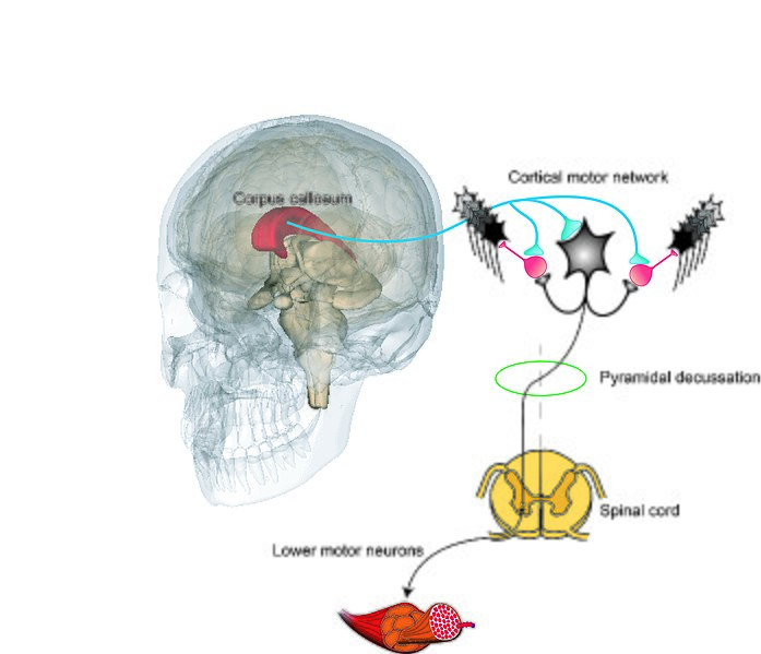
Functional unit of callosal influence
Summary[edit]
| Description |
English: The principal functional unit of callosal influence comprises a facilitatory centre and a depressing peripheral zone, that together shape the influence of converging inputs to pyramidal neurons. This organisation serves to sculpt and focus the output of neural circuits. In this schematic, cells that generate excitatory postsynaptic potentials (EPSPs) are represented in black. Callosal projections (excitatory) are represented in cyan. Inhibitory interneurons are represented in red. An inhibitory influence upon pyramidal tract cells in the surround region of the opposite hemisphere may arise from either local feed-forward inhibitory circuits or recurrent inhibitory circuits. Although the patterns of inhibition elicited by callosal projections are in part an emergent property of these local networks, the actuating excitatory drive (from the opposite hemisphere) is necessarily also selective and highly differentiated. A key assumption is that such inter-hemispheric interactions are reciprocal, and thus integrative.
The original artwork contains elements derived from the following sources: https://upload.wikimedia.org/wikipedia/commons/f/f3/Muscle_fascicles_and_cells.png; https://commons.wikimedia.org/wiki/File:Corpus_callosum.gif. |
| Date | |
| Source | Own work |
| Author | Rgcarson |
Licensing[edit]
I, the copyright holder of this work, hereby publish it under the following license:
This file is licensed under the Creative Commons Attribution-Share Alike 4.0 International license.
- You are free:
- to share – to copy, distribute and transmit the work
- to remix – to adapt the work
- Under the following conditions:
- attribution – You must give appropriate credit, provide a link to the license, and indicate if changes were made. You may do so in any reasonable manner, but not in any way that suggests the licensor endorses you or your use.
- share alike – If you remix, transform, or build upon the material, you must distribute your contributions under the same or compatible license as the original.
File history
Click on a date/time to view the file as it appeared at that time.
| Date/Time | Thumbnail | Dimensions | User | Comment | |
|---|---|---|---|---|---|
| current | 13:40, 31 May 2020 | | 4,763 × 4,096 (5.05 MB) | Rgcarson (talk | contribs) | Magnifying glass removed; corticospinal tract relationship to spinal cord clarified; annotations added |
| 12:03, 3 May 2020 |  | 4,529 × 4,686 (3.87 MB) | Rgcarson (talk | contribs) | Uploaded own work with UploadWizard |
You cannot overwrite this file.
File usage on Commons
There are no pages that use this file.