File:Crowded cytosol.png
Jump to navigation
Jump to search
Size of this preview: 696 × 600 pixels. Other resolutions: 279 × 240 pixels | 557 × 480 pixels | 892 × 768 pixels | 1,189 × 1,024 pixels | 2,308 × 1,988 pixels.
Original file (2,308 × 1,988 pixels, file size: 9.72 MB, MIME type: image/png)
Captions
Captions
Add a one-line explanation of what this file represents
Summary[edit]
| Description |
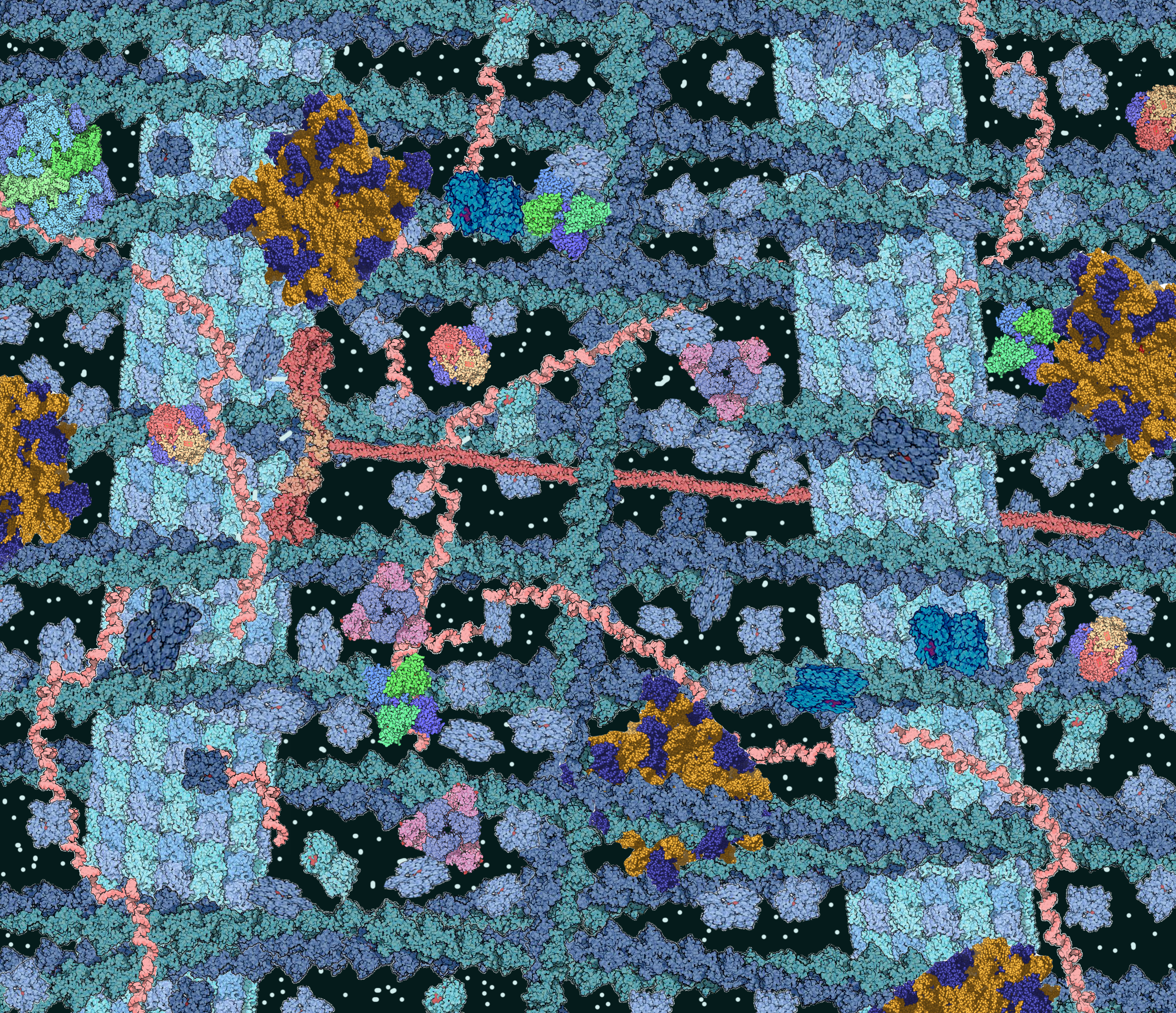
English: Picture of cytosol, showing microtubules (light blue), actin filaments (dark blue), ribosomes (yellow and purple), soluble proteins (light blue), kinesin (red), small molecules (white) and RNA (pink). |
| Source | Own work by uploader, based upon simlar illustrations in Goodsell DS (June 1991). "Inside a living cell". Trends Biochem. Sci. 16 (6): 203–6. DOI:10.1016/0968-0004(91)90083-8. PMID 1891800. |
| Author | TimVickers |
Licensing[edit]
| I, the copyright holder of this work, release this work into the public domain. This applies worldwide. In some countries this may not be legally possible; if so: I grant anyone the right to use this work for any purpose, without any conditions, unless such conditions are required by law. |
File history
Click on a date/time to view the file as it appeared at that time.
| Date/Time | Thumbnail | Dimensions | User | Comment | |
|---|---|---|---|---|---|
| current | 16:11, 11 September 2008 | | 2,308 × 1,988 (9.72 MB) | TimVickers (talk | contribs) | {{Information |Description={{en|1=Picture of cytosol, showing microtubules, actin filaments, ribosomes, soluble proteins and RNA}} |Source=Own work by uploader, collage of PD images from Category:PDB.org's Molecules of the Month by David S. Goodsell |
| 02:04, 11 September 2008 |  | 2,308 × 1,988 (10.68 MB) | TimVickers (talk | contribs) | {{Information |Description={{en|1=Picture of cytosol, showing microtubules, actin filaments, ribosomes, soluble proteins and RNA}} |Source=Own work by uploader |Author=TimVickers |Date= |Permission= |other_versions= }} <!--{{ImageUplo |
You cannot overwrite this file.
File usage on Commons
The following page uses this file:
File usage on other wikis
The following other wikis use this file:
- Usage on ar.wikipedia.org
- Usage on be.wikipedia.org
- Usage on bg.wikipedia.org
- Usage on bn.wikipedia.org
- Usage on bs.wikipedia.org
- Usage on ca.wikipedia.org
- Usage on cs.wikipedia.org
- Usage on cy.wikipedia.org
- Usage on de.wikipedia.org
- Usage on en.wikipedia.org
- Usage on en.wiktionary.org
- Usage on es.wikipedia.org
- Usage on fa.wikipedia.org
- Usage on ga.wikipedia.org
- Usage on gl.wikipedia.org
- Usage on he.wikipedia.org
- Usage on hy.wikipedia.org
- Usage on id.wikipedia.org
- Usage on it.wikipedia.org
- Usage on ja.wikipedia.org
- Usage on ko.wikipedia.org
- Usage on ml.wikipedia.org
- Usage on pl.wikipedia.org
- Usage on pt.wikipedia.org
- Usage on ru.wikipedia.org
- Usage on sh.wikipedia.org
- Usage on sr.wikipedia.org
- Usage on th.wikipedia.org
- Usage on tl.wikipedia.org
- Usage on tr.wikipedia.org
- Usage on uk.wikipedia.org
- Usage on www.wikidata.org
- Usage on zh.wikipedia.org