File:Craniopharyngioma2.jpg
Jump to navigation
Jump to search

Size of this preview: 405 × 600 pixels. Other resolutions: 162 × 240 pixels | 324 × 480 pixels | 518 × 768 pixels | 1,200 × 1,777 pixels.
Original file (1,200 × 1,777 pixels, file size: 261 KB, MIME type: image/jpeg)
Captions
Captions
Add a one-line explanation of what this file represents
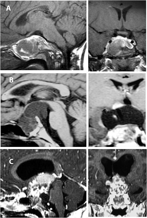
| Description | Enhanced T1 weighted MRI's of craniopharyngiomas. a. Apredominantly solid sellar/supra sellar tumour that is discrete fromthe hypothalamus (grade 0). b. A predominantly cystic sellar/suprasellar tumour that is distorting but not invading the hypothalamus (grade 1). c. A predominantly solid sellar/supra sellar tumour. The hypothalamus is not visible because of tumour invasion (grade 2). |
| Date | |
| Source | Matthew R Garnett, Stéphanie Puget, Jacques Grill, Christian Sainte-Rose. Craniopharyngioma. Orphanet Journal of Rare Diseases. 2, 18. 2007. PMID 17425791 |
| Author | see above |
| Permission (Reusing this file) |
http://www.biomedcentral.com/info/about/license |
This file is licensed under the Creative Commons Attribution 2.0 Generic license.
- You are free:
- to share – to copy, distribute and transmit the work
- to remix – to adapt the work
- Under the following conditions:
- attribution – You must give appropriate credit, provide a link to the license, and indicate if changes were made. You may do so in any reasonable manner, but not in any way that suggests the licensor endorses you or your use.
File history
Click on a date/time to view the file as it appeared at that time.
| Date/Time | Thumbnail | Dimensions | User | Comment | |
|---|---|---|---|---|---|
| current | 12:36, 6 August 2007 | | 1,200 × 1,777 (261 KB) | Filip em (talk | contribs) | {{Information |Description=Enhanced T1 weighted MRI's of craniopharyngiomas. a. Apredominantly solid sellar/supra sellar tumour that is discrete fromthe hypothalamus (grade 0). b. A predominantly cystic sellar/suprasellar tumour that is distorting but not |
You cannot overwrite this file.
File usage on Commons
The following page uses this file:
File usage on other wikis
The following other wikis use this file:
- Usage on ar.wikipedia.org
- Usage on en.wikipedia.org
- Usage on fa.wikipedia.org
- Usage on it.wikipedia.org
- Usage on nl.wikipedia.org
- Usage on pl.wikipedia.org
- Usage on tr.wikipedia.org