File:Classification and dissection of the insula.png
Jump to navigation
Jump to search

Size of this preview: 800 × 527 pixels. Other resolutions: 320 × 211 pixels | 640 × 422 pixels | 1,024 × 675 pixels | 1,280 × 843 pixels | 1,935 × 1,275 pixels.
Original file (1,935 × 1,275 pixels, file size: 2.38 MB, MIME type: image/png)
Captions
Captions
Add a one-line explanation of what this file represents
Summary[edit]
| Description |
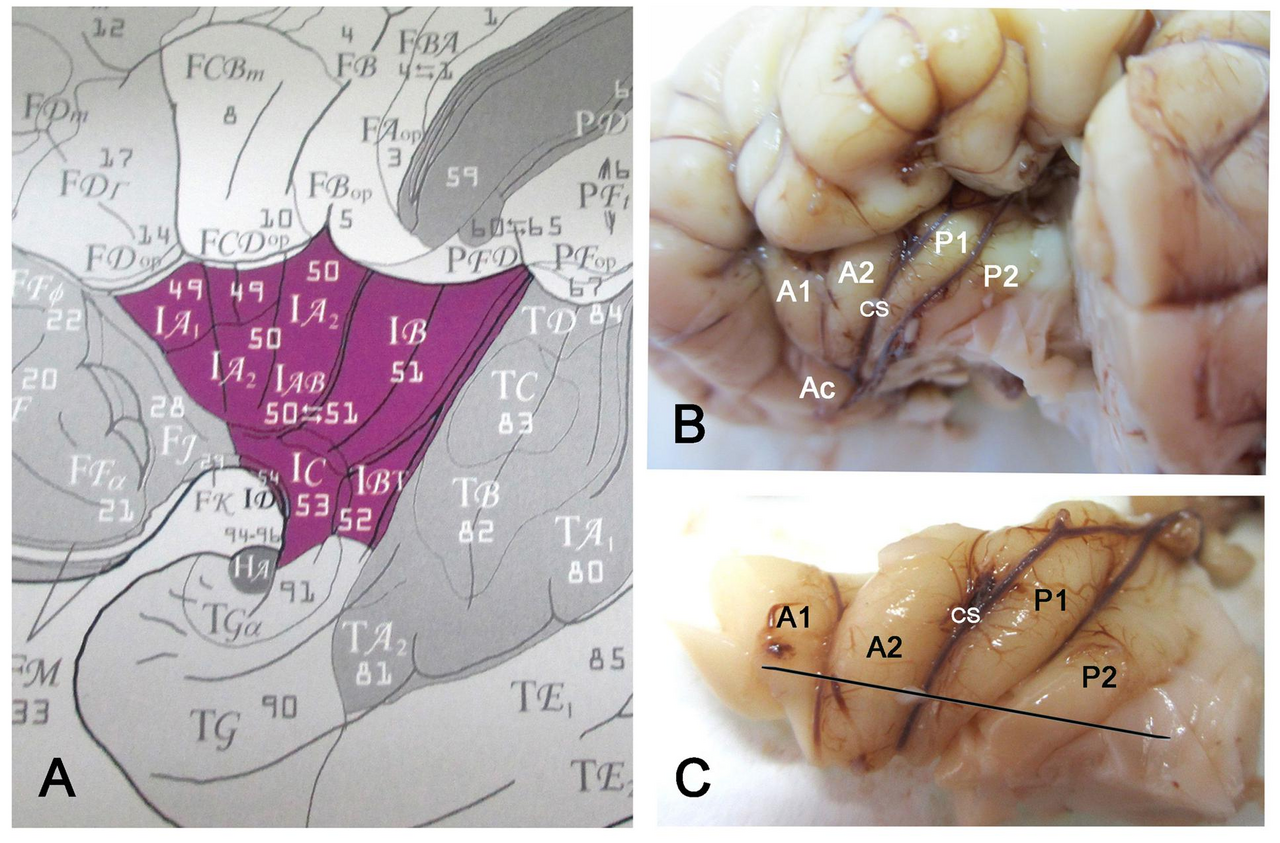
English: (A) Classification of the insular areas of Von Economo and Koskinas (1925). IA, anterior agranular, IB: posterior granular, IAB intermediate dysgranular areas. (B) Left insular lobe of a newborn infant (40 GW) after removing the anterior temporal pole. A1, A2, anterior short gyri; Ac: accessory short gyrus; P1, P2, posterior long gyri; CS: central sulcus of the insula. (C) Dissection of the insula in B. The line indicates the plane of section. |
| Date | Published: 05 December 2017 |
| Source | González-Arnay E, González-Gómez M and Meyer G (2017) A Radial Glia Fascicle Leads Principal Neurons from the Pallial-Subpallial Boundary into the Developing Human Insula. Front. Neuroanat. 11:111. https://doi.org/10.3389/fnana.2017.00111 |
| Author | Emilio González-Arnay, Miriam González-Gómez and Gundela Meyer |
Licensing[edit]
This file is licensed under the Creative Commons Attribution 4.0 International license.
- You are free:
- to share – to copy, distribute and transmit the work
- to remix – to adapt the work
- Under the following conditions:
- attribution – You must give appropriate credit, provide a link to the license, and indicate if changes were made. You may do so in any reasonable manner, but not in any way that suggests the licensor endorses you or your use.
File history
Click on a date/time to view the file as it appeared at that time.
| Date/Time | Thumbnail | Dimensions | User | Comment | |
|---|---|---|---|---|---|
| current | 09:36, 2 June 2019 | | 1,935 × 1,275 (2.38 MB) | Was a bee (talk | contribs) | {{Information |Description={{en|1=(A) Classification of the insular areas of Von Economo and Koskinas (1925). IA, anterior agranular, IB: posterior granular, IAB intermediate dysgranular areas. (B) Left insular lobe of a newborn infant (40 GW) after removing the anterior temporal pole. A1, A2, anterior short gyri; Ac: accessory short gyrus; P1, P2, posterior long gyri; CS: central sulcus of the insula. (C) Dissection of the insula in B. The line indicates the plane of section.}} |Source=Gonzá... |
You cannot overwrite this file.
File usage on Commons
There are no pages that use this file.