File:Bilirubin-from-xtal-1978-3D-balls.png
Jump to navigation
Jump to search
Size of this preview: 738 × 599 pixels. Other resolutions: 296 × 240 pixels | 591 × 480 pixels | 946 × 768 pixels | 1,261 × 1,024 pixels | 2,000 × 1,624 pixels.
Original file (2,000 × 1,624 pixels, file size: 595 KB, MIME type: image/png)
Captions
Captions
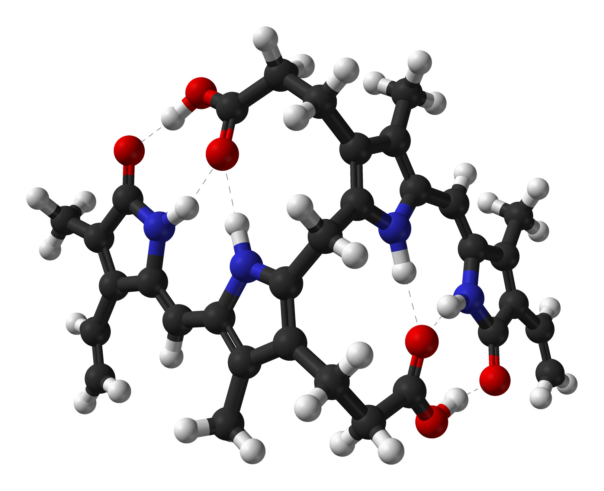
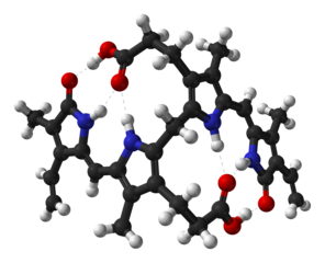
Bilirubin ball-and-stick model
| Description |
Ball-and-stick model of the bilirubin molecule as found in the crystal structure. Dashed lines are intramolecular hydrogen bonds. Colour code:
Structure by X-ray crystallography from Proc. R. Soc. Lond. B (1978) 202, 249-268. Image generated in Accelrys DS Visualizer. |
||
| Date | |||
| Source | Own work | ||
| Author | Ben Mills | ||
| Permission (Reusing this file) |
|
File history
Click on a date/time to view the file as it appeared at that time.
| Date/Time | Thumbnail | Dimensions | User | Comment | |
|---|---|---|---|---|---|
| current | 16:38, 5 February 2011 | | 2,000 × 1,624 (595 KB) | Benjah-bmm27 (talk | contribs) | {{Information |Description = Ball-and-stick model of the bilirubin molecule as found in the crystal structure. Dashed lines are intramolecular hydrogen bonds. '''Colour code:''' * Carbon, C: grey-black * Hydrogen, H: white * Nitrogen, |
You cannot overwrite this file.
File usage on Commons
The following 2 pages use this file:
File usage on other wikis
The following other wikis use this file:
- Usage on ar.wikipedia.org
- Usage on ary.wikipedia.org
- Usage on be.wikipedia.org
- Usage on bn.wikipedia.org
- Usage on ca.wikipedia.org
- Usage on ckb.wikipedia.org
- Usage on en.wikipedia.org
- Usage on es.wikipedia.org
- Usage on fa.wikipedia.org
- Usage on ga.wikipedia.org
- Usage on gl.wikipedia.org
- Usage on hu.wikipedia.org
- Usage on it.wikipedia.org
- Usage on ka.wikipedia.org
- Usage on ko.wikipedia.org
- Usage on lv.wikipedia.org
- Usage on mk.wikipedia.org
- Usage on ml.wikipedia.org
- Usage on pl.wikipedia.org
- Usage on pnb.wikipedia.org
- Usage on ro.wikipedia.org
- Usage on ru.wikipedia.org
- Usage on sh.wikipedia.org
- Usage on sr.wikipedia.org
- Usage on ta.wikipedia.org
- Usage on tg.wikipedia.org
- Usage on th.wikipedia.org
- Usage on tr.wikipedia.org
- Usage on ur.wikipedia.org
- Usage on vi.wikipedia.org
- Usage on www.wikidata.org