File:3D rendered CT of abdominal aortic branches and kidneys.svg
Jump to navigation
Jump to search

Size of this PNG preview of this SVG file: 507 × 352 pixels. Other resolutions: 320 × 222 pixels | 640 × 444 pixels | 1,024 × 711 pixels | 1,280 × 889 pixels | 2,560 × 1,777 pixels.
Original file (SVG file, nominally 507 × 352 pixels, file size: 546 KB)
Captions
Captions
Add a one-line explanation of what this file represents
Summary[edit]
| Description |
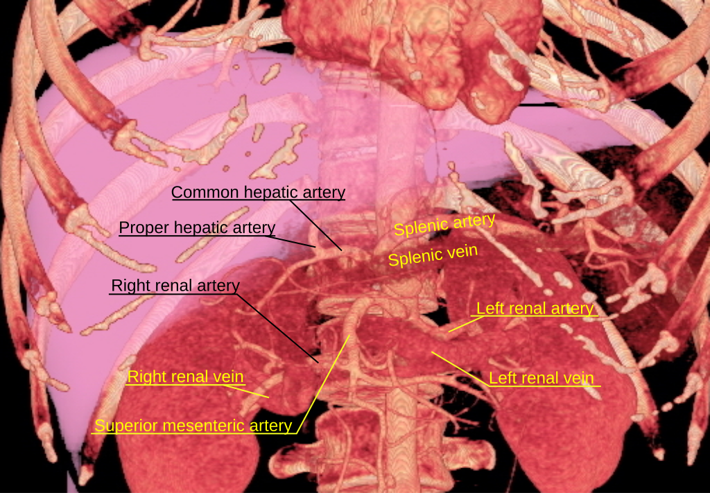
English: 3D rendered computed tomography of abdominal aortic branches and kidneys. Inferior vena cava is not visible, however. |
| Date | |
| Source | LDLT volume measure.jpg. |
| Author | Mikael Häggström from original image created and uploaded by Dr. I-Chen Tsai (Wikipedia:User:Sillyduck) |
| Other versions |
 |
Licensing[edit]

|
Permission is granted to copy, distribute and/or modify this document under the terms of the GNU Free Documentation License, Version 1.2 or any later version published by the Free Software Foundation; with no Invariant Sections, no Front-Cover Texts, and no Back-Cover Texts. A copy of the license is included in the section entitled GNU Free Documentation License. |
This file is licensed under the Creative Commons Attribution 3.0 Unported license.
- You are free:
- to share – to copy, distribute and transmit the work
- to remix – to adapt the work
- Under the following conditions:
- attribution – You must give appropriate credit, provide a link to the license, and indicate if changes were made. You may do so in any reasonable manner, but not in any way that suggests the licensor endorses you or your use.
File history
Click on a date/time to view the file as it appeared at that time.
| Date/Time | Thumbnail | Dimensions | User | Comment | |
|---|---|---|---|---|---|
| current | 18:10, 22 October 2010 | | 507 × 352 (546 KB) | Mikael Häggström (talk | contribs) | {{Information |Description={{en|1=f}} |Source={{own}} |Author=Mikael Häggström |Date=f |Permission= |other_versions= }} |
You cannot overwrite this file.
File usage on Commons
The following page uses this file:
File usage on other wikis
The following other wikis use this file:
- Usage on ar.wikipedia.org
- Usage on az.wikipedia.org
- Usage on bs.wikipedia.org
- Usage on en.wikipedia.org
- Usage on es.wikipedia.org
- Usage on eu.wikipedia.org
- Usage on fa.wikipedia.org
- Usage on fr.wikipedia.org
- Usage on ja.wikipedia.org
- Usage on ko.wikipedia.org
- Usage on ks.wikipedia.org
- Usage on la.wikipedia.org
- Usage on nl.wikipedia.org
- Usage on ro.wikipedia.org
- Usage on www.wikidata.org