File:197-Zika Virus-ZikaVirus.tif
Jump to navigation
Jump to search

Size of this JPG preview of this TIF file: 467 × 600 pixels. Other resolutions: 187 × 240 pixels | 374 × 480 pixels | 598 × 768 pixels | 797 × 1,024 pixels | 2,487 × 3,193 pixels.
Original file (2,487 × 3,193 pixels, file size: 12.88 MB, MIME type: image/tiff)
Captions
Captions
Add a one-line explanation of what this file represents
Summary[edit]
| Description |
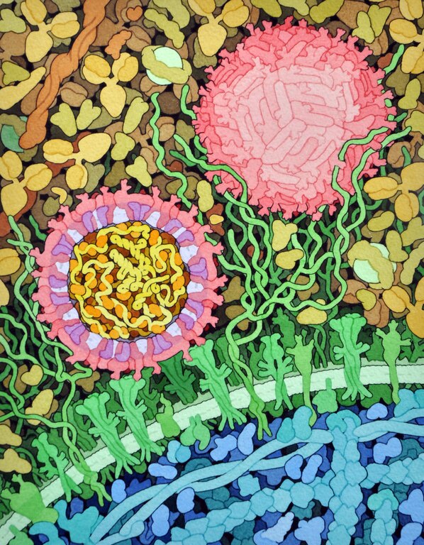
English: Space-fill drawing of the outside of one Zika virus particle, and a cross-section through another as it interacts with a cell. The two main proteins of the viral envelope, the envelope proteins and membrane proteins, are shown in red and purple respectively. The lipid membrane of the envelope is shown in light lavender.The capsid proteins, in orange, are shown interacting with the RNA genome, in yellow, at the center of the virus. The cell-surface receptor proteins are in green, the cytoskeleton in blue, and blood plasma proteins in gold. Drawn by David Goodsell. |
| Date | |
| Source | RCSB Molecule of the Month 197, June 2016 |
| Author | David Goodsell |
Licensing[edit]
This file is licensed under the Creative Commons Attribution 4.0 International license.
- You are free:
- to share – to copy, distribute and transmit the work
- to remix – to adapt the work
- Under the following conditions:
- attribution – You must give appropriate credit, provide a link to the license, and indicate if changes were made. You may do so in any reasonable manner, but not in any way that suggests the licensor endorses you or your use.
File history
Click on a date/time to view the file as it appeared at that time.
| Date/Time | Thumbnail | Dimensions | User | Comment | |
|---|---|---|---|---|---|
| current | 18:23, 5 June 2016 |  | 2,487 × 3,193 (12.88 MB) | Dcrjsr (talk | contribs) | User created page with UploadWizard |
You cannot overwrite this file.
File usage on Commons
The following page uses this file:
File usage on other wikis
The following other wikis use this file:
- Usage on en.wikipedia.org
- Talk:Alan Turing
- Talk:Amino acid
- Talk:Apoptosis
- Talk:Bacteria
- Talk:Biochemistry
- Talk:Outline of biology
- Talk:Biotechnology
- Talk:Cell (biology)
- Talk:Cladistics
- Talk:Citric acid cycle
- Talk:Cell cycle
- Talk:Molecular diffusion
- Talk:Major depressive disorder
- Talk:Dolly (sheep)
- Talk:Extremophile
- Talk:Endosymbiont
- Talk:Evolutionary tree
- Talk:Endoplasmic reticulum
- Talk:Enzyme
- Talk:Functional group
- Talk:Fat
- Talk:Genetically modified organism
- Talk:Guanine
- Talk:Genetic programming
- Talk:Genetic code
- Talk:Glycolysis
- Talk:Glucose
- Talk:Hemoglobin
- Talk:Human cloning
- Talk:Insulin
- Talk:Interferon
- Talk:Lipid
- Talk:List of algorithms
- Talk:Mutagenesis
- Talk:MARCKS protein
- Talk:Mitosis
- Talk:Microevolution
- Talk:Miller–Urey experiment
- Talk:Neuron
- Talk:Prion
- Talk:Retrovirus
- Talk:RNA world
- Talk:Stephen Jay Gould
- Talk:Stem cell
- Talk:Toluene
- Talk:The Bell Curve
- Talk:Vitamin
- Talk:Vitamin C
- Talk:Uracil
- Talk:Cytochrome
View more global usage of this file.