File:0311 Pancreatic Cells Micrograph.jpg

Original file (822 × 390 pixels, file size: 219 KB, MIME type: image/jpeg)
Captions
Captions
Summary[edit]
| Description0311 Pancreatic Cells Micrograph.jpg |
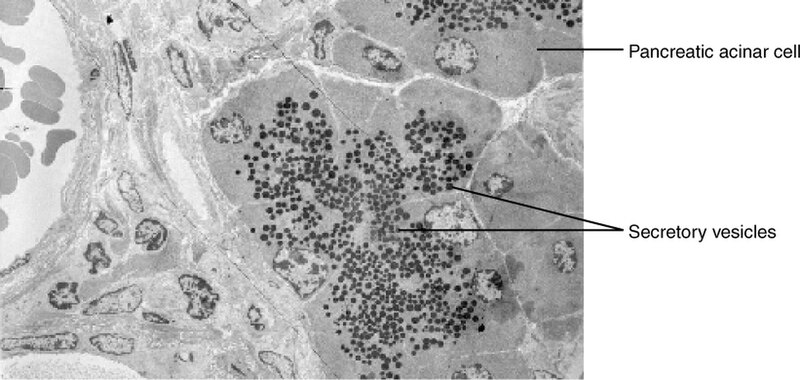
Caption: The pancreatic acinar cells produce and secrete many enzymes that digest food. The tiny black granules in this electron micrograph are secretory vesicles filled with enzymes that will be exported from the cells via exocytosis. LM × 2900. (Micrograph provided by the Regents of University of Michigan Medical School © 2012) |
| Date | |
| Source | https://cnx.org/contents/FPtK1zmh@8.25:fEI3C8Ot@10/Preface |
| Author | OpenStax |
| Other versions |
|
Licensing[edit]
- You are free:
- to share – to copy, distribute and transmit the work
- to remix – to adapt the work
- Under the following conditions:
- attribution – You must give appropriate credit, provide a link to the license, and indicate if changes were made. You may do so in any reasonable manner, but not in any way that suggests the licensor endorses you or your use.
File history
Click on a date/time to view the file as it appeared at that time.
| Date/Time | Thumbnail | Dimensions | User | Comment | |
|---|---|---|---|---|---|
| current | 16:58, 3 July 2016 |  | 822 × 390 (219 KB) | CFCF (talk | contribs) | |
| 16:35, 3 July 2016 |  | 822 × 390 (219 KB) | CFCF (talk | contribs) | ||
| 09:40, 13 December 2013 |  | 822 × 390 (219 KB) | CFCF (talk | contribs) | User created page with UploadWizard |
You cannot overwrite this file.
File usage on Commons
The following 3 pages use this file:
Metadata
This file contains additional information such as Exif metadata which may have been added by the digital camera, scanner, or software program used to create or digitize it. If the file has been modified from its original state, some details such as the timestamp may not fully reflect those of the original file. The timestamp is only as accurate as the clock in the camera, and it may be completely wrong.
| Software used | Illustrator |
|---|---|
| Date and time of digitizing | 23:21, 2 April 2013 |
| File change date and time | 07:21, 2 April 2013 |
| Date metadata was last modified | 23:21, 2 April 2013 |
| Copyright status | Copyright status not set |

