File:Virus Replication large.svg

Original file (SVG file, nominally 925 × 852 pixels, file size: 310 KB)
Captions
Captions
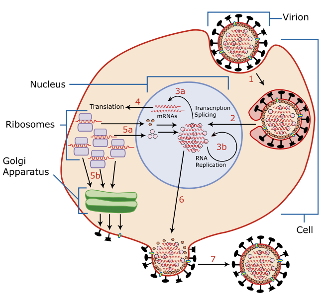
| Description | A diagram of influenza viral cell invasion and replication. | ||||||||
| Date | |||||||||
| Source | Scaled up from Image:Virus Replication.svg by User:YK Times, who redrew from w:Image:Virusreplication.png using Adobe Illustrator. Based on Scheme of Influenza A virus replication, NCBI, artwork by David Hobson, August 6, 2002 | ||||||||
| Author | User:YK Times | ||||||||
| Permission (Reusing this file) |
I, the copyright holder of this work, hereby publish it under the following licenses:
This file is licensed under the Creative Commons Attribution-Share Alike 2.5 Generic, 2.0 Generic and 1.0 Generic license.
You may select the license of your choice. |
||||||||
| Other versions |
|

.png)


Summary[edit]
From Scheme of Influenza A virus replication (NCBI):
"A virion attaches to the host cell membrane using Hemagglutinin(HA) and enters the cytoplasm by receptor-mediated endocytosis (STEP 1), thereby forming an endosome. A cellular trypsin-like enzyme cleaves HA into products HA1 and HA2 (not shown). HA2 promotes fusion of the virus envelope and the endosome membranes. A minor virus envelope protein M2 acts as a ion channel making the inside of the virion more acidic. As a result, the major envelope protein M1 dissociates from the nucleocapsid and vRNAs are translocated into the nucleus (STEP 2) via interaction between NP and cellular transport machinery. In the nucleus, the viral polymerase complexes transcribe (STEP 3a) and replicate (STEP 3b) the vRNAs. Newly synthesized mRNAs migrate to cytoplasm (STEP 4) where they are translated. Posttranslational processing of HA, Neuraminidase(NA), and M2 includes transportation via Golgi apparatus to the cell membrane (STEP 5b). NP, M1, NS1 (nonstructural regulatory protein - not shown) and NEP (nuclear export protein, a minor virion component - not shown) move to the nucleus (STEP 5a) and bind freshly synthesized copies of vRNAs. The newly formed nucleocapsids migrate into the cytoplasm in a NEP-dependent process and eventually interact via protein M1 with a region of the cell membrane where HA, NA and M2 have been inserted (STEP 6). Then the newly synthesized virions bud from infected cell (STEP 7). NA destroys the sialic acid moiety of cellular receptors, thereby releasing the progeny virions.
| Annotations | This image is annotated: View the annotations at Commons |
File history
Click on a date/time to view the file as it appeared at that time.
| Date/Time | Thumbnail | Dimensions | User | Comment | |
|---|---|---|---|---|---|
| current | 17:26, 16 March 2008 | | 925 × 852 (310 KB) | Photohound (talk | contribs) | {{Information |Description=A diagram of influenza viral cell invasion and replication. |Source=Scaled up from Image:Virus Replication.svg by User:YK Times, who redrew from w:Image:Virusreplication.png using Adobe Illustrator. |Date=March 5, 2 |
You cannot overwrite this file.
File usage on Commons
The following 2 pages use this file:
File usage on other wikis
The following other wikis use this file:
- Usage on ar.wikipedia.org
- Usage on da.wikipedia.org
- Usage on de.wikipedia.org
- Usage on de.wikibooks.org
- Usage on el.wiktionary.org
- Usage on en.wikipedia.org
- Usage on es.wikipedia.org
- Usage on eu.wikipedia.org
- Usage on hu.wikipedia.org
- Usage on hu.wikibooks.org
- Usage on mk.wikipedia.org
- Usage on ms.wikipedia.org
- Usage on pt.wikipedia.org
- Usage on ru.wikipedia.org
- Usage on th.wikipedia.org
- Usage on uk.wikipedia.org
- Usage on zh.wikipedia.org