File:Virus Replication.svg

Original file (SVG file, nominally 462 × 426 pixels, file size: 205 KB)
Captions
Captions
Summary[edit]
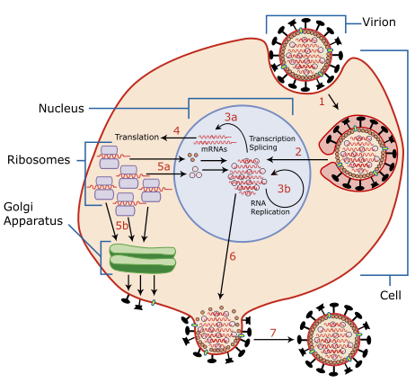
| Description | A diagram of influenza viral cell invasion and replication. |
| Date | |
| Source | Redrawn from w:Image:Virusreplication.png using Adobe Illustrator. |
| Author | User:YK Times |
| Other versions |
 |


|
This SVG file contains embedded text that can be translated into your language, using any capable SVG editor, text editor or the SVG Translate tool. For more information see: About translating SVG files. |
Description from Scheme of Influenza A virus replication (NCBI): "A virion attaches to the host cell membrane via HA and enters the cytoplasm by receptor-mediated endocytosis (STEP 1), thereby forming an endosome. A cellular trypsin-like enzyme cleaves HA into products HA1 and HA2 (not shown). HA2 promotes fusion of the virus envelope and the endosome membranes. A minor virus envelope protein M2 acts as a ion channel thereby making the inside of the virion more acidic. As a result, the major envelope protein M1 dissociates from the nucleocapsid and vRNPs are translocated into the nucleus (STEP 2) via interaction between NP and cellular transport machinery. In the nucleus, the viral polymerase complexes transcribe (STEP 3a) and replicate (STEP 3b) the vRNAs. Newly synthesized mRNAs migrate to cytoplasm (STEP 4) where they are translated. Posttranslational processing of HA, NA, and M2 includes transportation via Golgi apparatus to the cell membrane (STEP 5b). NP, M1, NS1 (nonstructural regulatory protein - not shown) and NEP (nuclear export protein, a minor virion component - not shown) move to the nucleus (STEP 5a) where they bind freshly synthesized copies of vRNAs. The newly formed nucleocapsids migrate into the cytoplasm in a NEP-dependent process and eventually interact via M1 with a region of the cell membrane where HA, NA and M2 have been inserted (STEP 6). Then the newly synthesized virions bud from infected cell (STEP 7). NA destroys the sialic acid moiety of cellular receptors, thereby releasing the progeny virions."
Licensing[edit]

|
Permission is granted to copy, distribute and/or modify this document under the terms of the GNU Free Documentation License, Version 1.2 or any later version published by the Free Software Foundation; with no Invariant Sections, no Front-Cover Texts, and no Back-Cover Texts. A copy of the license is included in the section entitled GNU Free Documentation License. |
| This file is licensed under the Creative Commons Attribution-Share Alike 3.0 Unported license. | ||
| ||
| This licensing tag was added to this file as part of the GFDL licensing update. |
- You are free:
- to share – to copy, distribute and transmit the work
- to remix – to adapt the work
- Under the following conditions:
- attribution – You must give appropriate credit, provide a link to the license, and indicate if changes were made. You may do so in any reasonable manner, but not in any way that suggests the licensor endorses you or your use.
- share alike – If you remix, transform, or build upon the material, you must distribute your contributions under the same or compatible license as the original.
| Annotations | This image is annotated: View the annotations at Commons |
File history
Click on a date/time to view the file as it appeared at that time.
| Date/Time | Thumbnail | Dimensions | User | Comment | |
|---|---|---|---|---|---|
| current | 02:54, 6 March 2007 | | 462 × 426 (205 KB) | YK Times (talk | contribs) | {{Information |Description=A diagram of influenza viral cell invasion and replication. |Source=Redrawn from w:Image:Virusreplication.png using Adobe Illustrator. |Date=March 5, 2007 |Author= User:YK Times |Permission= |other_versions=[[:w:Image:V |
You cannot overwrite this file.
File usage on Commons
The following 5 pages use this file:
.png)
File usage on other wikis
The following other wikis use this file:
- Usage on bg.wikipedia.org
- Usage on bn.wikipedia.org
- Usage on br.wikipedia.org
- Usage on ca.wikipedia.org
- Usage on cs.wikipedia.org
- Usage on da.wikipedia.org
- Usage on de.wikipedia.org
- Usage on el.wikipedia.org
- Usage on en.wikipedia.org
- Orthomyxoviridae
- Amantadine
- Viral replication
- Viral life cycle
- Viral entry
- File:Virusreplication.png
- User:YK Times/Graphic Lab/examples
- Wikipedia:Graphics Lab/Images to improve/Archive/Mar 2007
- Viral shedding
- Template:Influenza virus life cycle
- Viral neuraminidase
- Tilapia tilapinevirus
- User:Zoe.gum/sandbox
- Wikipedia talk:WikiProject Viruses/Archive 4
- User:Anicm1/sandbox
- Usage on en.wikibooks.org
- Usage on es.wikipedia.org
- Usage on fa.wikipedia.org
- Usage on fr.wikipedia.org
- Usage on hy.wikipedia.org
- Usage on id.wikipedia.org
- Usage on it.wikipedia.org
- Usage on ja.wikipedia.org
- Usage on kk.wikipedia.org
- Usage on ko.wikipedia.org
View more global usage of this file.