File:Transverse section at the level of the abdomen. human (01).jpg
Jump to navigation
Jump to search
Size of this preview: 800 × 285 pixels. Other resolutions: 320 × 114 pixels | 640 × 228 pixels | 1,024 × 364 pixels | 1,280 × 455 pixels | 4,036 × 1,436 pixels.
Original file (4,036 × 1,436 pixels, file size: 2.91 MB, MIME type: image/jpeg)
Captions
Captions
Add a one-line explanation of what this file represents
Summary[edit]
.jpg&action=edit§ion=1)
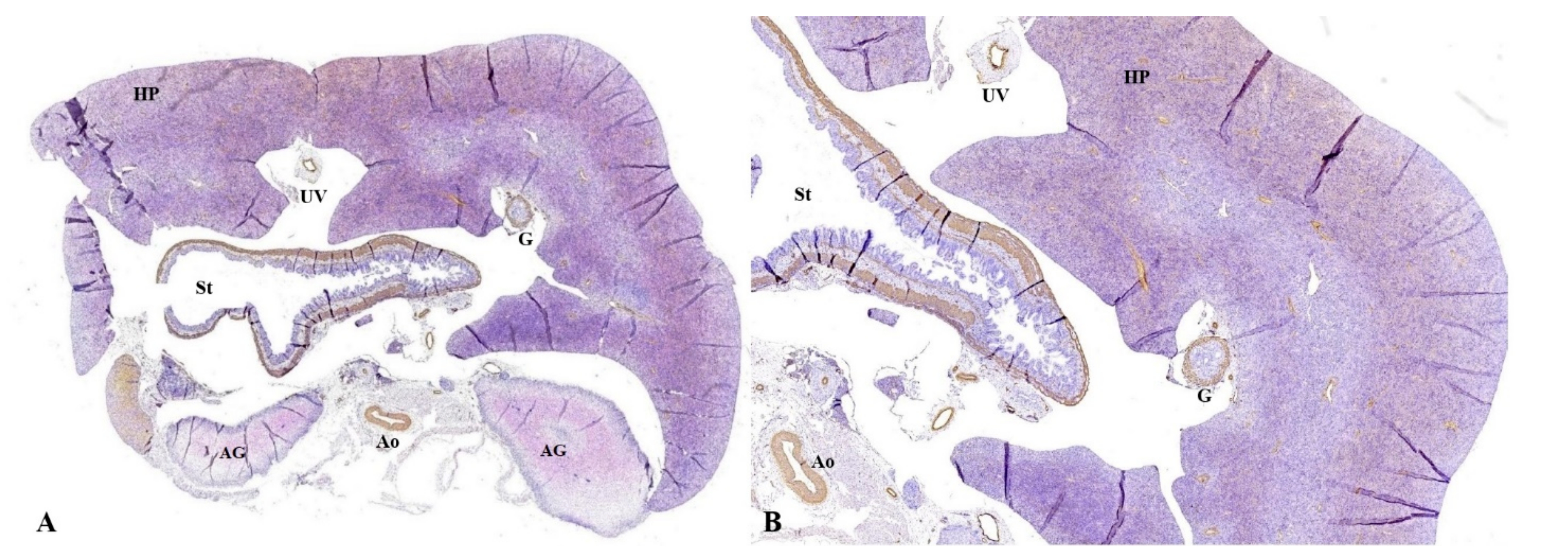
| Description | Figure 10. Transverse section at the level of the abdomen. (A): The liver parenchyma (HP) located anteriorly, the disorganized hepatocyte cords, the muscular tunics of the blood vessels immunolabeled with brown in both the liver parenchyma and the great hepatic vessels, the muscular and the middle tunic of the stomach (St), muscular tunic of the aorta (Ao); (B): the hepatic parenchyma, disorganized hepatocyte cords, muscle tunics of the blood vessels immunolabeled with brown in both the hepatic parenchyma (HP) and the great hepatic vessels are observed. Moreover, the muscular and the middle tunic of the stomach (St), muscular tunic of the aorta (Ao) are identified. Immunohistochemical staining with anti-αSMA antibody. αSMA: alpha actin smooth muscle, HP: hepatic parenchyma, UV: umbilical vein, G: gallbladder, St: stomach, AG: adrenal glands, Ao: Aorta, Sp: spleen. |
| Date | |
| Source | Feasibility of Fetal Portal Venous System Ultrasound Assessment at the FT Anomaly Scan. Diagnostics 2022, 12, 361. https://doi.org/10.3390/diagnostics12020361 |
| Author | Nagy, R.D.; Ruican, D.; Zorilă, G.-L.; Istrate-Ofiţeru, A.-M.; Badiu, A.M.; Iliescu, D.G. |
Open acces
Licensing[edit]
.jpg&action=edit§ion=2)
This file is licensed under the Creative Commons Attribution 4.0 International license.
- You are free:
- to share – to copy, distribute and transmit the work
- to remix – to adapt the work
- Under the following conditions:
- attribution – You must give appropriate credit, provide a link to the license, and indicate if changes were made. You may do so in any reasonable manner, but not in any way that suggests the licensor endorses you or your use.

|
This file, which was originally posted to an external website, has not yet been reviewed by an administrator or reviewer to confirm that the above license is valid. See Category:License review needed for further instructions.
|
File history
Click on a date/time to view the file as it appeared at that time.
| Date/Time | Thumbnail | Dimensions | User | Comment | |
|---|---|---|---|---|---|
| current | 21:34, 15 February 2024 | 4,036 × 1,436 (2.91 MB) | Rasbak (talk | contribs) | {{Information |description=Figure 10. Transverse section at the level of the abdomen. (A): The liver parenchyma (HP) located anteriorly, the disorganized hepatocyte cords, the muscular tunics of the blood vessels immunolabeled with brown in both the liver parenchyma and the great hepatic vessels, the muscular and the middle tunic of the stomach (St), muscular tunic of the aorta (Ao); (B): the hepatic parenchyma, disorganized hepatocyte cords, muscle tunics of the blood vessels immunolabeled w... |
You cannot overwrite this file.
File usage on Commons
There are no pages that use this file.
.jpg&oldid=852168379)