File:Pre- and post-central gyrus, right hemisphere.jpg
Jump to navigation
Jump to search

Size of this preview: 380 × 599 pixels. Other resolutions: 152 × 240 pixels | 304 × 480 pixels | 884 × 1,394 pixels.
Original file (884 × 1,394 pixels, file size: 1.13 MB, MIME type: image/jpeg)
Captions
Captions
Add a one-line explanation of what this file represents
Summary[edit]
| Description |
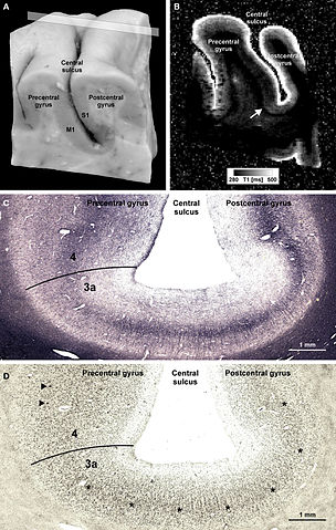
English: Pre- and post-central gyrus, right hemisphere. Microstructural 7-T MR mapping in post-mortem brains. (A) Tissue block (pre- and post-central gyrus, right hemisphere) from a post-mortem human brain (female, 61 years, died of pulmonary failure and chronic obstructive pulmonary disease, autopsy performed with informed consent of the patient’s relatives, post-mortem interval before fixation 24 h, fixed in 4% formalin for 2 months) prior to MR scanning and histological processing. M1 = primary motor cortex in the posterior wall of the precentral gyrus, S1 = primary somatosensory cortex in the anterior wall of the post-central gyrus. (B) Quantitative T1 map of the tissue block [for plane of sectioning see rectangle in (A)]. MP2RAGE sequence at 7 T, voxel size (0.33 mm)3, 32 averages, acquisition time 3 h 50 min, surrounding medium: Fomblin (Solvay Solexis, Bollate, Italy). Arrow indicates a sharp change in T1 contrast at the base of the precentral gyrus that matches a change in the myelo- and cytoarchitectonic pattern [cf. (C,D)]. (C,D) Frozen sections (30 μm) from a corresponding position of the same block stained for myelin basic protein [rat monoclonal antibody, avidin–biotin–peroxidase complex (ABC) method, chromogen: DAB and ammonium nickel(II) sulfate (C)] and cell bodies. Micrographs show the fundus of the central sulcus [same orientation as in (A,B)]. The drop in T1 values at the base of the precentral gyrus coincides with an increase in myelin basic protein immunostaining [line in (C)]. In an accompanying section stained for cell bodies, this position is characterized by an increase in gray matter thickness, a disappearing inner granular layer (asterisks), and emerging giant pyramidal (Betz) cells (arrowheads). This transition [lines in (C,D)] corresponds to the border between area 3a (somatosensory cortex) and area 4 (primary motor cortex). |
| Date | Published online: 2011-02-18 |
| Source | Geyer S, Weiss M, Reimann K, Lohmann G and Turner R (2011) Microstructural parcellation of the human cerebral cortex – from Brodmann’s post-mortem map to in vivo mapping with high-field magnetic resonance imaging. Front. Hum. Neurosci. 5:19. doi: 10.3389/fnhum.2011.00019 http://journal.frontiersin.org/article/10.3389/fnhum.2011.00019/full |
| Author | Geyer S, Weiss M, Reimann K, Lohmann G and Turner R |
Licensing[edit]
This file is licensed under the Creative Commons Attribution 3.0 Unported license.
- You are free:
- to share – to copy, distribute and transmit the work
- to remix – to adapt the work
- Under the following conditions:
- attribution – You must give appropriate credit, provide a link to the license, and indicate if changes were made. You may do so in any reasonable manner, but not in any way that suggests the licensor endorses you or your use.
File history
Click on a date/time to view the file as it appeared at that time.
| Date/Time | Thumbnail | Dimensions | User | Comment | |
|---|---|---|---|---|---|
| current | 21:54, 9 September 2015 | | 884 × 1,394 (1.13 MB) | Was a bee (talk | contribs) | {{Information |Description={{en|1=Pre- and post-central gyrus, right hemisphere. Microstructural 7-T MR mapping in post-mortem brains. (A) Tissue block (pre- and post-central gyrus, right hemisphere) from a post-mortem human brain (female, 61 years, di... |
You cannot overwrite this file.
File usage on Commons
The following page uses this file:
File usage on other wikis
The following other wikis use this file:
- Usage on es.wikipedia.org