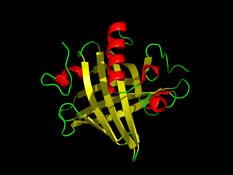
File:Mup1 PDB 1i04.jpg
From Wikimedia Commons, the free media repository
Jump to navigation
Jump to search

Size of this preview: 800 × 600 pixels. Other resolutions: 320 × 240 pixels | 640 × 480 pixels | 1,024 × 768 pixels | 1,280 × 960 pixels.
Original file (1,280 × 960 pixels, file size: 207 KB, MIME type: image/jpeg)
File information
Structured data
Captions
Captions
Add a one-line explanation of what this file represents
Summary[edit]

|
This file was moved to Wikimedia Commons from en.wikipedia using a bot script. All source information is still present. It requires review. Additionally, there may be errors in any or all of the information fields; information on this file should not be considered reliable and the file should not be used until it has been reviewed and any needed corrections have been made. Once the review has been completed, this template should be removed. For details about this file, see below. Check now! |
| DescriptionMup1 PDB 1i04.jpg |
English: Tertiary structure of mouse major urinary protein I. Resolved from PDB:1i04. The protein has eight beta sheets (yellow) arranged in a beta barrel open at one end, with alpha helices (red) at both the amino- and carboxyl termini. |
| Date | 12/03/08 |
| Source | I created this work entirely by myself, using AISMIG |
| Author | Rockpocket |
Licensing[edit]
| Public domainPublic domainfalsefalse |
| This work has been released into the public domain by its author, Rockpocket at English Wikipedia. This applies worldwide. In some countries this may not be legally possible; if so: Rockpocket grants anyone the right to use this work for any purpose, without any conditions, unless such conditions are required by law.Public domainPublic domainfalsefalse |
Original upload log[edit]
Transferred from en.wikipedia to Commons by Bobamnertiopsis using CommonsHelper.
The original description page was here. All following user names refer to en.wikipedia.
- 2008-12-03 08:41 Rockpocket 1280×960× (212381 bytes) {{Information |Description= Structure of mouse major urinary protein I. Resolved from PDB:1i04 |Source=I created this work entirely by myself. |Date= |Author=~~~ |other_versions= }}
File history
Click on a date/time to view the file as it appeared at that time.
| Date/Time | Thumbnail | Dimensions | User | Comment | |
|---|---|---|---|---|---|
| current | 22:26, 2 February 2010 |  | 1,280 × 960 (207 KB) | File Upload Bot (Magnus Manske) (talk | contribs) | {{BotMoveToCommons|en.wikipedia|year={{subst:CURRENTYEAR}}|month={{subst:CURRENTMONTHNAME}}|day={{subst:CURRENTDAY}}}} {{Information |Description={{en|en:Tertiary structure of mouse major urinary protein I. Resolved from [http://www.rcsb.org/pdb/ex |
You cannot overwrite this file.
File usage on Commons
There are no pages that use this file.
Metadata
This file contains additional information such as Exif metadata which may have been added by the digital camera, scanner, or software program used to create or digitize it. If the file has been modified from its original state, some details such as the timestamp may not fully reflect those of the original file. The timestamp is only as accurate as the clock in the camera, and it may be completely wrong.
| _error | 0 |
|---|