File:Medial view of a cadaver dissection of the gastrocnemius–soleus junction.png
Jump to navigation
Jump to search

Size of this preview: 416 × 599 pixels. Other resolutions: 167 × 240 pixels | 333 × 480 pixels | 858 × 1,235 pixels.
Original file (858 × 1,235 pixels, file size: 1.09 MB, MIME type: image/png)
Captions
Captions
Add a one-line explanation of what this file represents
Summary[edit]
| Description |
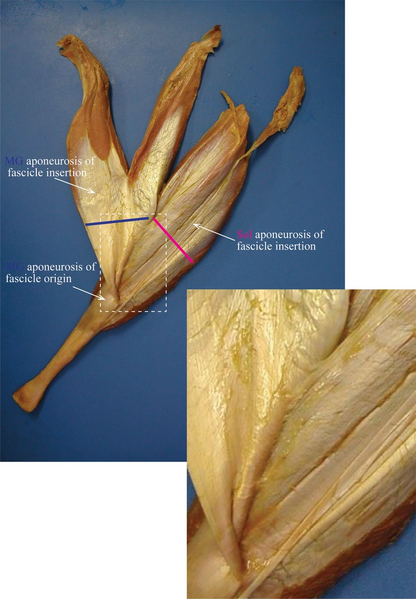
English: Medial view of a cadaver dissection of the gastrocnemius–soleus junction proximal to the Achilles tendon. Skin, subcutaneous tissue, and superficial fascia are removed. The gastrocnemius and soleus (Sol) muscle fibers are behind each aponeurosis. The gastrocnemius aponeurosis continues for a variable distance inferior to the distal ends of the medial and lateral heads of gastrocnemius to reach its line of attachment to the aponeurosis of the Sol fascicle insertion. The attachment between the aponeuroses of the medial gastrocnemius (MG) and Sol fascicles insertion enlarges on the bottom‐right corner. Blue and pink lines correspond to locations where the myotendinous junction occurred and where the membrane thickness of the aponeurosis was measured using microscopy. |
| Date | Published online 2013 Nov 7 |
| Source |
Kinugasa R, Oda T, Komatsu T, Edgerton VR, Sinha S. Interaponeurosis shear strain modulates behavior of myotendinous junction of the human triceps surae. Physiol Rep. 2013;1(6):e00147. doi:10.1002/phy2.147 https://physoc.onlinelibrary.wiley.com/doi/full/10.1002/phy2.147 |
| Author | Ryuta Kinugasa,1,2,3 Toshiaki Oda,2,4 Toshihiko Komatsu,5 V Reggie Edgerton,6,7,8 and Shantanu Sinha |
Licensing[edit]
This file is licensed under the Creative Commons Attribution 4.0 International license.
- You are free:
- to share – to copy, distribute and transmit the work
- to remix – to adapt the work
- Under the following conditions:
- attribution – You must give appropriate credit, provide a link to the license, and indicate if changes were made. You may do so in any reasonable manner, but not in any way that suggests the licensor endorses you or your use.
File history
Click on a date/time to view the file as it appeared at that time.
| Date/Time | Thumbnail | Dimensions | User | Comment | |
|---|---|---|---|---|---|
| current | 22:09, 4 April 2020 | | 858 × 1,235 (1.09 MB) | Was a bee (talk | contribs) | {{Information |Description={{en|1=Medial view of a cadaver dissection of the gastrocnemius–soleus junction proximal to the Achilles tendon. Skin, subcutaneous tissue, and superficial fascia are removed. The gastrocnemius and soleus (Sol) muscle fibers are behind each aponeurosis. The gastrocnemius aponeurosis continues for a variable distance inferior to the distal ends of the medial and lateral heads of gastrocnemius to reach its line of attachment to the aponeurosis of the Sol fascicle inse... |
You cannot overwrite this file.
File usage on Commons
The following page uses this file: