File:Low vs high resolution hemoglobin detail.jpg
Jump to navigation
Jump to search
Size of this preview: 800 × 600 pixels. Other resolutions: 320 × 240 pixels | 640 × 480 pixels | 1,024 × 768 pixels.
Original file (1,024 × 768 pixels, file size: 300 KB, MIME type: image/jpeg)
Captions
Captions
Add a one-line explanation of what this file represents
Summary[edit]
| Description |
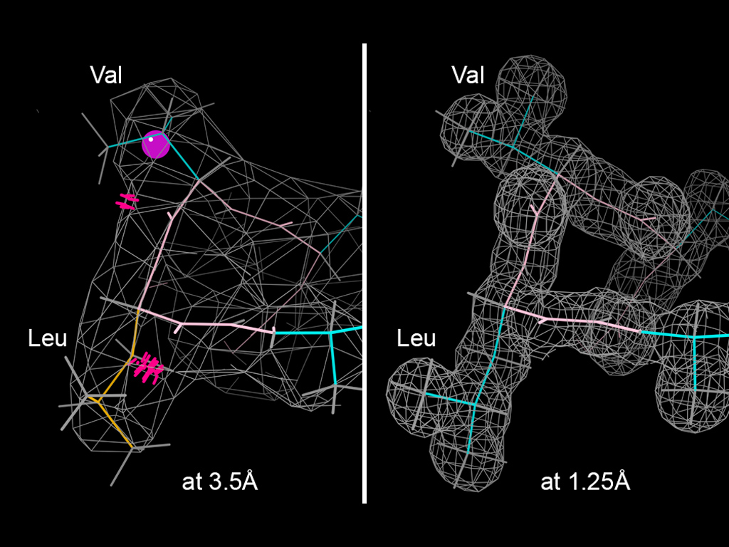
English: Comparison of low vs high resolution electron density for the same detail of crystal structure on a helix in hemoglobin (2QLS at 3.5Å vs 2DN2 at 1.25Å). Not surprisingly, the low-resolution density is hard to fit accurately, and it shows bad steric overlaps ("clashes", marked with red spikes) and incorrect sidechain conformations. Validation diagnosis in MolProbity, display in KiNG. |
| Date | |
| Source | Own work |
| Author | Dcrjsr |
Licensing[edit]
This file is licensed under the Creative Commons Attribution 3.0 Unported license.
- You are free:
- to share – to copy, distribute and transmit the work
- to remix – to adapt the work
- Under the following conditions:
- attribution – You must give appropriate credit, provide a link to the license, and indicate if changes were made. You may do so in any reasonable manner, but not in any way that suggests the licensor endorses you or your use.
File history
Click on a date/time to view the file as it appeared at that time.
| Date/Time | Thumbnail | Dimensions | User | Comment | |
|---|---|---|---|---|---|
| current | 17:34, 15 May 2011 | | 1,024 × 768 (300 KB) | Dcrjsr (talk | contribs) | {{Information |Description ={{en|1=Comparison of low vs high resolution electron density for the same detail of crystal structure on a helix in hemoglobin (2QLS at 3.5Å vs 2DN2 at 1.25Å). Not surprisingly, the low-resolution density is hard to fit a |
You cannot overwrite this file.
File usage on Commons
The following page uses this file:
File usage on other wikis
The following other wikis use this file:
- Usage on en.wikipedia.org