File:Leghemoglobin 1FSL.png
Jump to navigation
Jump to search

Size of this preview: 775 × 600 pixels. Other resolutions: 310 × 240 pixels | 620 × 480 pixels | 992 × 768 pixels | 1,280 × 991 pixels | 1,620 × 1,254 pixels.
Original file (1,620 × 1,254 pixels, file size: 602 KB, MIME type: image/png)
Captions
Captions
Add a one-line explanation of what this file represents
| Description |
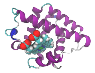
English: Ribbon model of soybean leghemoglobin a, with heme as balls, after PDB 1FSL. Ref.: Ellis PJ, Appleby CA, Guss JM, Hunter WN, Ollis DL, Freeman HC (May 1997). "Structure of ferric soybean leghemoglobin a nicotinate at 2.3 A resolution". Acta Crystallogr. D Biol. Crystallogr. 53 (Pt 3): 302–10. DOI:10.1107/S0907444997000292. PMID 15299933.
This image was created with VMD. |
||
| Date | |||
| Source | Own work | ||
| Author |
Ayacop Please flattr this |
||
| Permission (Reusing this file) |
|
File history
Click on a date/time to view the file as it appeared at that time.
| Date/Time | Thumbnail | Dimensions | User | Comment | |
|---|---|---|---|---|---|
| current | 07:55, 20 December 2009 | | 1,620 × 1,254 (602 KB) | Ayacop (talk | contribs) | {{Information |Description={{en|1=Ribbon model of soybean leghemoglobin a, with heme as balls, after PDB 1FSL. Ref.: {{cite journal |author=Ellis PJ, Appleby CA, Guss JM, Hunter WN, Ollis DL, Freeman HC |title=Structure of ferric soybean leghemoglobin a n |
You cannot overwrite this file.
File usage on Commons
There are no pages that use this file.
File usage on other wikis
The following other wikis use this file:
- Usage on ar.wikipedia.org
- Usage on da.wikipedia.org
- Usage on de.wikipedia.org
- Usage on fa.wikipedia.org
- Usage on fr.wikipedia.org
- Usage on gl.wikipedia.org
- Usage on he.wikipedia.org
- Usage on mk.wikipedia.org
- Usage on ml.wikipedia.org
- Usage on nl.wikipedia.org
- Usage on pl.wikipedia.org
- Usage on pt.wikipedia.org
- Usage on ru.wikipedia.org
- Usage on zh.wikipedia.org