File:Illustration of scapholunate interosseous ligament anatomy.png
From Wikimedia Commons, the free media repository
Jump to navigation
Jump to search

Size of this preview: 500 × 599 pixels. Other resolutions: 200 × 240 pixels | 564 × 676 pixels.
Original file (564 × 676 pixels, file size: 252 KB, MIME type: image/png)
File information
Structured data
Captions
Captions
Add a one-line explanation of what this file represents
Summary[edit]
| DescriptionIllustration of scapholunate interosseous ligament anatomy.png |
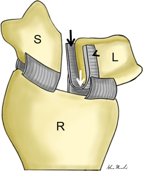
English: Illustration of scapholunate interosseous ligament anatomy. The drawing depicts a slightly oblique, coronal view of the distal radius (R), scaphoid (S) and lunate (L). The scapholunate ligament has been transected to demonstrate its three distinct parts, which include the dorsal (arrowhead), membranous (white arrow) and volar (black arrow) components. Note that the dorsal component is the thickest |
| Date | |
| Source | Tischler, B.T., Diaz, L.E., Murakami, A.M. et al. Scapholunate advanced collapse: a pictorial review. Insights Imaging 5, 407–417 (2014). https://doi.org/10.1007/s13244-014-0337-1 |
| Author | Brian T. Tischler, Luis E. Diaz, Akira M. Murakami, Frank W. Roemer, Ajay R. Goud, William F. Arndt III & Ali Guermazi |
Licensing[edit]
This file is licensed under the Creative Commons Attribution 4.0 International license.
- You are free:
- to share – to copy, distribute and transmit the work
- to remix – to adapt the work
- Under the following conditions:
- attribution – You must give appropriate credit, provide a link to the license, and indicate if changes were made. You may do so in any reasonable manner, but not in any way that suggests the licensor endorses you or your use.
File history
Click on a date/time to view the file as it appeared at that time.
| Date/Time | Thumbnail | Dimensions | User | Comment | |
|---|---|---|---|---|---|
| current | 14:28, 10 April 2021 |  | 564 × 676 (252 KB) | Balkanique (talk | contribs) | Uploaded a work by Brian T. Tischler, Luis E. Diaz, Akira M. Murakami, Frank W. Roemer, Ajay R. Goud, William F. Arndt III & Ali Guermazi from Tischler, B.T., Diaz, L.E., Murakami, A.M. et al. Scapholunate advanced collapse: a pictorial review. Insights Imaging 5, 407–417 (2014). https://doi.org/10.1007/s13244-014-0337-1 with UploadWizard |
You cannot overwrite this file.
File usage on Commons
The following page uses this file:
Hidden category: