File:Human skeletal muscle tissue 3 - TEM.jpg

Original file (2,400 × 1,745 pixels, file size: 1.4 MB, MIME type: image/jpeg)
Captions
Captions
Summary[edit]
| Description |
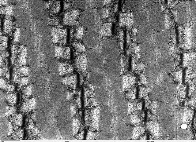
Transmission electron microscope image of a thin longitudinal section cut through an area of human skeletal muscle tissue. Image of muscle sarcomeres shows distinct banding pattern: the darker bands are called A bands(the A band includes a lighter central zone, called the H band), and the lighter bands are called I bands. Each I band is bisected by a dark transverse line called the Z-line). Paired mitochondria are on either side of the electron opaque Z-line. The Z-Line marks the longitudinal extent of a sarcomere unit. JEOL 100CX TEM |
| Source | |
| Author | Louisa Howard |
| Permission (Reusing this file) |
PD |
Licensing[edit]
| This work has been released into the public domain by its author, Louisa Howard. This applies worldwide. In some countries this may not be legally possible; if so: Louisa Howard grants anyone the right to use this work for any purpose, without any conditions, unless such conditions are required by law.
|
File history
Click on a date/time to view the file as it appeared at that time.
| Date/Time | Thumbnail | Dimensions | User | Comment | |
|---|---|---|---|---|---|
| current | 15:02, 7 October 2006 | | 2,400 × 1,745 (1.4 MB) | Patho (talk | contribs) | {{Information |Description=Transmission electron microscope image of a thin longitudinal section cut through an area of human skeletal muscle tissue. Image of muscle sarcomeres shows distinct banding pattern: the darker bands are called A bands(the A band |
You cannot overwrite this file.
File usage on Commons
There are no pages that use this file.
File usage on other wikis
The following other wikis use this file:
- Usage on de.wikibooks.org