File:Human-insulin-hexamer-3D-ribbons.png
Jump to navigation
Jump to search
Size of this preview: 613 × 599 pixels. Other resolutions: 246 × 240 pixels | 491 × 480 pixels | 786 × 768 pixels | 1,100 × 1,075 pixels.
Original file (1,100 × 1,075 pixels, file size: 524 KB, MIME type: image/png)
Captions
Captions
Add a one-line explanation of what this file represents
Summary[edit]
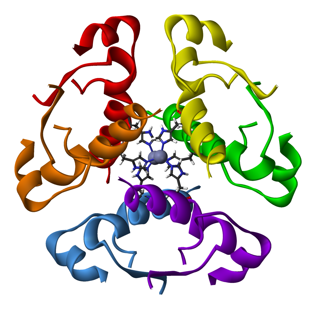
| Description | Ribbon diagram of the 2-Zn insulin hexamer. The insulin monomer is the active hormone, but the hexamer is the storage form. Each of the 6 monomers is shown in a different color, and the 6 histidine side-chains that bind the hexamer-stabilizing zincs are detailed as stick-figures. The spirals are alpha helices and the arrows are beta strands. | ||
| Source | Own work | ||
| Author | Benjah-bmm27 | ||
| Permission (Reusing this file) |
|
|
File history
Click on a date/time to view the file as it appeared at that time.
| Date/Time | Thumbnail | Dimensions | User | Comment | |
|---|---|---|---|---|---|
| current | 20:38, 22 April 2007 | | 1,100 × 1,075 (524 KB) | Benjah-bmm27 (talk | contribs) | {{PD-user|Benjah-bmm27}} Category:Insulin |
You cannot overwrite this file.
File usage on Commons
There are no pages that use this file.
File usage on other wikis
The following other wikis use this file:
- Usage on an.wikipedia.org
- Usage on ast.wikipedia.org
- Usage on bn.wikibooks.org
- Usage on ca.wikipedia.org
- Usage on de.wikipedia.org
- Usage on en.wikipedia.org
- Usage on en.wikibooks.org
- Usage on es.wikipedia.org
- Usage on fa.wikipedia.org
- Usage on id.wikipedia.org
- Usage on pt.wikipedia.org
- Usage on tr.wikipedia.org
- Usage on uk.wikipedia.org