File:Hsv encephalitis.jpg
Jump to navigation
Jump to search
No higher resolution available.
Hsv_encephalitis.jpg (480 × 512 pixels, file size: 88 KB, MIME type: image/jpeg)
Captions
Captions
Add a one-line explanation of what this file represents
| Description |
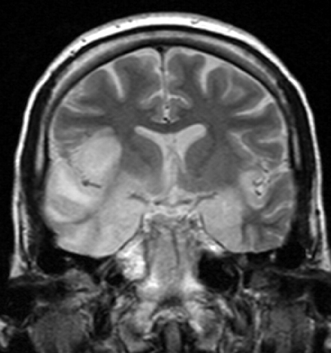
"This 33 year-old female patient presented with agitation, confusion, mutism, and fever. This coronal T2-weighted MR image shows high signal in the temporal lobes including hippocampal formations and parahippogampal gyrae, insulae, and right inferior frontal gyrus. The right gyrus rectus and the columns of the fornices were also involved (not shown). There was no associated haemorrhage or enhancement. There was moderate mass-effect. Lumbar puncture was performed - herpes simplex virus DNA was not shown on polymerase chain reaction. A brain biopsy was performed and the histology was consistent with encephalitis. PCR was repeated on the biopsy specimen and was positive for HSV type I." (dr Dawes) |
| Date | |
| Source | http://www.radpod.org/2007/03/24/herpes-simplex-encephalitis/ |
| Author | dr Laughlin Dawes |
This file is licensed under the Creative Commons Attribution 3.0 Unported license.
- You are free:
- to share – to copy, distribute and transmit the work
- to remix – to adapt the work
- Under the following conditions:
- attribution – You must give appropriate credit, provide a link to the license, and indicate if changes were made. You may do so in any reasonable manner, but not in any way that suggests the licensor endorses you or your use.
File history
Click on a date/time to view the file as it appeared at that time.
| Date/Time | Thumbnail | Dimensions | User | Comment | |
|---|---|---|---|---|---|
| current | 10:44, 15 May 2008 | | 480 × 512 (88 KB) | Filip em (talk | contribs) | {{Information |Description=This 33 year-old female patient presented with agitation, confusion, mutism, and fever. This coronal T2-weighted MR image shows high signal in the temporal lobes including hippocampal formations and parahippogampal gyrae, insula |
You cannot overwrite this file.
File usage on Commons
The following page uses this file:
File usage on other wikis
The following other wikis use this file:
- Usage on af.wikipedia.org
- Usage on an.wikipedia.org
- Usage on ar.wikipedia.org
- Usage on as.wikipedia.org
- Usage on be.wikipedia.org
- Usage on bg.wikipedia.org
- Usage on bn.wikipedia.org
- Usage on ca.wikipedia.org
- Usage on ckb.wikipedia.org
- Usage on da.wikipedia.org
- Usage on de.wikipedia.org
- Usage on de.wikibooks.org
- Usage on el.wikipedia.org
- Usage on en.wikipedia.org
- Usage on es.wikipedia.org
- Usage on et.wikipedia.org
- Usage on eu.wikipedia.org
- Usage on fa.wikipedia.org
- Usage on fr.wikipedia.org
- Usage on ga.wikipedia.org
- Usage on gl.wikipedia.org
- Usage on he.wikipedia.org
- Usage on hi.wikipedia.org
- Usage on hu.wikipedia.org
- Usage on hy.wikipedia.org
- Usage on it.wikipedia.org
- Usage on ja.wikipedia.org
View more global usage of this file.