File:HindIII 2E52.png
Jump to navigation
Jump to search

Size of this preview: 539 × 600 pixels. Other resolutions: 216 × 240 pixels | 431 × 480 pixels | 690 × 768 pixels | 921 × 1,024 pixels | 1,496 × 1,664 pixels.
Original file (1,496 × 1,664 pixels, file size: 1.27 MB, MIME type: image/png)
Captions
Captions
Add a one-line explanation of what this file represents
| Description |
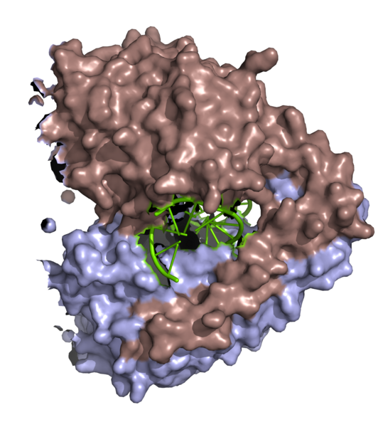
English: 3d surface model of HindIII dimer complexed with a DNA fragment from PDB 2E52. Ref.: Watanabe, N., Sato, C., Takasaki, Y., Tanaka, I. Crystal structural analysis of HindIII restriction endonuclease in complex with cognate DNA at 2.0 angstrom resolution to be published |
||
| Date | |||
| Source | adapted from http://www.pdb.org/pdb/files/2e52.pdb using PyMOL | ||
| Author | Ayacop | ||
| Permission (Reusing this file) |
|
||
| Other versions | http://www.ebi.ac.uk/pdbsum/2E52 |
File history
Click on a date/time to view the file as it appeared at that time.
| Date/Time | Thumbnail | Dimensions | User | Comment | |
|---|---|---|---|---|---|
| current | 17:59, 1 November 2008 | | 1,496 × 1,664 (1.27 MB) | Ayacop (talk | contribs) | {{Information |Description={{en|1=3d surface model of '''Hin'''dIII dimer complexed with a DNA fragment from PDB 2E52. Ref.: Watanabe, N., Sato, C., Takasaki, Y., Tanaka, I. Crystal structural analysis of HindIII restriction endonuclease in complex w |
You cannot overwrite this file.
File usage on Commons
There are no pages that use this file.
File usage on other wikis
The following other wikis use this file:
- Usage on de.wikipedia.org
- Usage on zh.wikipedia.org