File:Hepatic stellate cell (ito cell) 1476-5926-6-7-3-l.jpg
Jump to navigation
Jump to search

Size of this preview: 369 × 599 pixels. Other resolutions: 148 × 240 pixels | 423 × 687 pixels.
Original file (423 × 687 pixels, file size: 178 KB, MIME type: image/jpeg)
Captions
Captions
Add a one-line explanation of what this file represents
| Description |
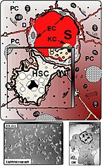
English: Schematic presentation of hepatic stellate cells (HSC) located in the vicinity of adjacent hepatocytes (PC) beneath the sinusoidal endothelial cells (EC). S – liver sinusoids; KC – Kupffer cells. Down left shows cultured HSC at light-microscopy, whereas at down right electron microscopy (EM) illustrates numerous fat vacuoles (L) in a HSC, in which retinoids are stored.
Gressner et al. Comparative Hepatology 2007 6:7 doi:10.1186/1476-5926-6-7 |
| Date | |
| Source | http://www.comparative-hepatology.com/content/6/1/7 |
| Author | Gressner et al. Comparative Hepatology 2007 6:7 doi:10.1186/1476-5926-6-7 |
| Permission (Reusing this file) |
© 2007 Gressner et al; licensee BioMed Central Ltd. This is an Open Access article: verbatim copying and redistribution of this article are permitted in all media for any purpose, provided this notice is preserved along with the article's original URL |
This file is licensed under the Creative Commons Attribution-Share Alike 3.0 Unported license.
- You are free:
- to share – to copy, distribute and transmit the work
- to remix – to adapt the work
- Under the following conditions:
- attribution – You must give appropriate credit, provide a link to the license, and indicate if changes were made. You may do so in any reasonable manner, but not in any way that suggests the licensor endorses you or your use.
- share alike – If you remix, transform, or build upon the material, you must distribute your contributions under the same or compatible license as the original.
File history
Click on a date/time to view the file as it appeared at that time.
| Date/Time | Thumbnail | Dimensions | User | Comment | |
|---|---|---|---|---|---|
| current | 12:18, 4 August 2008 | | 423 × 687 (178 KB) | CopperKettle (talk | contribs) | {{Information |Description={{en|1=Schematic presentation of hepatic stellate cells (HSC) located in the vicinity of adjacent hepatocytes (PC) beneath the sinusoidal endothelial cells (EC). S – liver sinusoids; KC – Kupffer cells. Down left shows cultu |
You cannot overwrite this file.
File usage on Commons
The following page uses this file:
File usage on other wikis
The following other wikis use this file:
- Usage on cs.wikipedia.org
- Usage on en.wikipedia.org
- Usage on es.wikipedia.org
- Usage on gl.wikipedia.org
- Usage on it.wikipedia.org
- Usage on ka.wikipedia.org
- Usage on pl.wikipedia.org
- Usage on pt.wikipedia.org
- Usage on ru.wikipedia.org
- Usage on sl.wikipedia.org
- Usage on www.wikidata.org
_1476-5926-6-7-3-l.jpg&oldid=595464979)