File:Hematopoiesis (human) diagram es.svg
Jump to navigation
Jump to search

Size of this PNG preview of this SVG file: 775 × 600 pixels. Other resolutions: 310 × 240 pixels | 620 × 480 pixels | 992 × 768 pixels | 1,280 × 991 pixels | 2,560 × 1,981 pixels | 1,416 × 1,096 pixels.
Original file (SVG file, nominally 1,416 × 1,096 pixels, file size: 4.79 MB)
Captions
Captions
Add a one-line explanation of what this file represents
Summary[edit]
| Description |
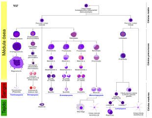
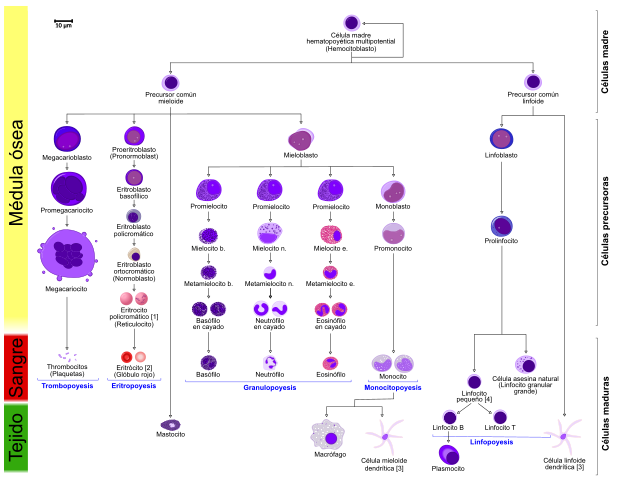
Español: Hematopoyesis humana
[1] El eritrocito policromático (reticulocito) de la derecha muestra su aspecto característico cuando se tiñe con azul de metileno o Azure B. [2] El eritrocito de la derecha es una representación más precisa de su apariencia en la realidad cuando se ve a través de un microscopio. [3] Otras células que surgen del monocito: osteoclastos, microglia (sistema nervioso central), células de Langerhans (epidermis), células de Kupffer (hígado). [4] Para mayor claridad, los linfocitos T y B se dividen para indicar mejor que la célula plasmática surge de la célula B. Tenga en cuenta que no hay diferencia en la apariencia de las células B y T, a menos que se aplique una tinción específica. |
| Date | |
| Source |
|
| Author |
Vector: and .jpg) - Reusing images - Conflicts of interest: None |
| Other versions |
[] This SVG[edit]
Other SVG[edit]
PNG[edit]
PNG with notes box[edit]
|
| SVG development | This oversized diagram was created with Inkscape. This diagram uses embedded text that can be easily translated using a text editor. |
_diagram_switch.svg&lang=en)
_diagram_switch.svg&lang=ca)
_diagram_switch.svg&lang=es)
_diagram_switch.svg&lang=gl)
_diagram_switch.svg&lang=is)
_diagram_switch.svg&lang=pt)
_diagram_switch.svg&lang=ru)
_diagram_switch.svg&lang=ja)
_diagram_switch.svg&lang=zh-cn)
_diagram.svg)
_diagram_fr.svg)
_diagram_is.png)

_diagram_zh.png)
_diagram.png)
_diagram_en.png)
_diagram-es.png)
_diagram_uk.png)
_diagram_es.svg&action=edit§ion=1)
_diagram_en.svg)
To modify the text of this drawing, read the content of this link.
Licensing[edit]
_diagram_es.svg&action=edit§ion=2)
I, the copyright holder of this work, hereby publish it under the following license:
This file is licensed under the Creative Commons Attribution-Share Alike 3.0 Unported license.
- You are free:
- to share – to copy, distribute and transmit the work
- to remix – to adapt the work
- Under the following conditions:
- attribution – You must give appropriate credit, provide a link to the license, and indicate if changes were made. You may do so in any reasonable manner, but not in any way that suggests the licensor endorses you or your use.
- share alike – If you remix, transform, or build upon the material, you must distribute your contributions under the same or compatible license as the original.
File history
Click on a date/time to view the file as it appeared at that time.
| Date/Time | Thumbnail | Dimensions | User | Comment | |
|---|---|---|---|---|---|
| current | 20:23, 19 July 2021 | | 1,416 × 1,096 (4.79 MB) | Jmarchn (talk | contribs) | Validation W3C |
| 13:19, 28 March 2021 |  | 1,416 × 1,096 (4.74 MB) | -sasha- (talk | contribs) | Dependiendo de la fuente se ponen uno o tres promielocitos, prefiero dejar tres | |
| 14:13, 20 March 2021 |  | 1,416 × 1,096 (4.42 MB) | -sasha- (talk | contribs) | Los promielocitos no presentan granulación secundaria y son todos iguales morfológicamente. Consultado en el atlas de histología de Ross | |
| 17:42, 21 December 2017 |  | 1,416 × 1,096 (4.5 MB) | Jmarchn (talk | contribs) | User created page with UploadWizard |
You cannot overwrite this file.
File usage on Commons
The following page uses this file:
_diagram_es.svg)
File usage on other wikis
The following other wikis use this file:
- Usage on es.wikipedia.org
_diagram_es.svg&oldid=680653889)