File:Heart kabuki syndrome.jpg
Jump to navigation
Jump to search

Size of this preview: 800 × 536 pixels. Other resolutions: 320 × 214 pixels | 640 × 429 pixels | 1,024 × 686 pixels | 1,200 × 804 pixels.
Original file (1,200 × 804 pixels, file size: 177 KB, MIME type: image/jpeg)
Captions
Captions
Add a one-line explanation of what this file represents
Summary[edit]
| Description |
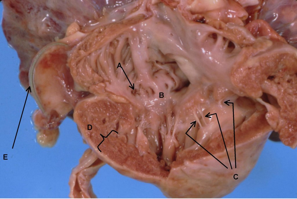
English: Anatomy of right ventricle and atrium. A. Dilated coronary sinus. B. Dysplastic tricuspid valve. C. Short thickened chordae tendinae almost implanted into papillary muscle. D. Right ventricular hypertrophy with ventricular wall thickness of 8 mm. E. Pacer wire. |
| Date | |
| Source | Shah M., Bogucki B., Mavers M., deMello DE., Knutsen A. Cardiac conduction abnormalities and congenital immunodeficiency in a child with Kabuki syndrome: case report.. BMC Med Genet. Jul 25;6, 28. 2005. doi:10.1186/1471-2350-6-28. PMID 16042804. |
| Author | see above |
| Permission (Reusing this file) |
[1] |
Licensing[edit]
This file is licensed under the Creative Commons Attribution 2.0 Generic license.
- You are free:
- to share – to copy, distribute and transmit the work
- to remix – to adapt the work
- Under the following conditions:
- attribution – You must give appropriate credit, provide a link to the license, and indicate if changes were made. You may do so in any reasonable manner, but not in any way that suggests the licensor endorses you or your use.
File history
Click on a date/time to view the file as it appeared at that time.
| Date/Time | Thumbnail | Dimensions | User | Comment | |
|---|---|---|---|---|---|
| current | 15:58, 17 June 2008 | | 1,200 × 804 (177 KB) | Filip em (talk | contribs) | {{Information |Description={{en|1=Anatomy of right ventricle and atrium. A. Dilated coronary sinus. B. Dysplastic tricuspid valve. C. Short thickened chordae tendinae almost implanted into papillary muscle. D. Right ventricular hypertrophy with ventricula |
You cannot overwrite this file.
File usage on Commons
There are no pages that use this file.
File usage on other wikis
The following other wikis use this file:
- Usage on pl.wikipedia.org
- Usage on pt.wikipedia.org